How to Detect FIP in Cats Without Ascites
Feline Infectious Peritonitis (FIP) is a deadly disease caused by a mutated feline coronavirus. While the classic presentation involves the accumulation of fluid within the abdominal cavity—ascites—some cats with FIP do not develop this hallmark symptom. Detecting FIP in these cases can be challenging but is crucial for early intervention and accurate diagnosis. This article explores various methods and considerations for diagnosing FIP in cats without ascites.
Understanding FIP without Ascites
FIP manifests primarily in two forms: the wet (effusive) and dry (non-effusive) forms. The wet form is characterized by fluid buildup in the abdomen or chest, easily detectable via physical examination and ultrasound. Conversely, the dry form lacks significant effusion and presents with more subtle signs, including granulomatous lesions in organs, neurological issues, or ocular abnormalities. Diagnosing FIP without ascites requires careful observation of clinical signs and a combination of diagnostic approaches.
Clinical Signs and History
A thorough clinical assessment forms the foundation for diagnosis. Cats with FIP often exhibit nonspecific symptoms such as weight loss, lethargy, fever, anorexia, and malaise. In the dry form, neurological signs—like ataxia or seizures—or ocular changes—such as uveitis—may predominate. A detailed history, including exposure to multi-cat environments and prior health status, provides valuable context.
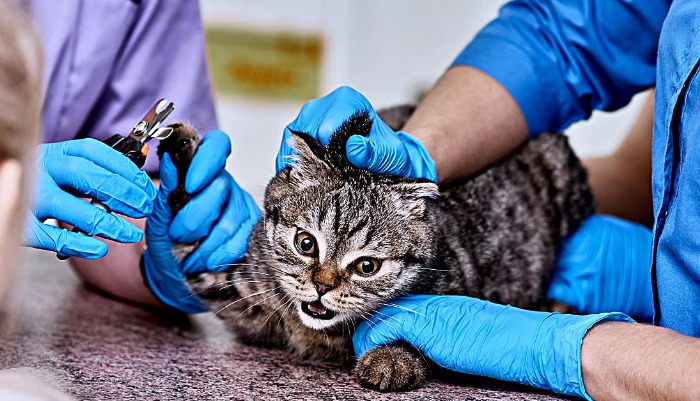
Physical Examination
During physical exams, subtle signs may be the only clues. Look for lymphadenopathy, jaundice, or abnormalities in the eyes or neurological examination. Although absence of ascites reduces suspicion, persistent signs of illness warrant further testing.
Laboratory Tests
Laboratory investigations are vital. Complete blood counts frequently reveal lymphopenia, neutrophilia, or anemia. Serum biochemistry tests may show elevated liver enzymes or hyperglobulinemia, reflecting systemic inflammation. However, these findings are nonspecific and must be combined with other diagnostic methods.
Serology and Antibody Tests
Serological tests detecting coronavirus antibodies indicate exposure but do not confirm FIP. High antibody titers suggest exposure but do not differentiate between benign infection and FIP. False positives and negatives are common, limiting their diagnostic utility alone.
Molecular Diagnostics: RT-PCR
Reverse Transcription Polymerase Chain Reaction (RT-PCR) assays detect coronavirus RNA in tissues or bodily fluids. While highly sensitive, they can produce false positives because they detect benign coronavirus strains, not the mutated FIP-causing form. Testing of tissue biopsies, particularly of affected organs, enhances specificity.
Histopathology and Tissue Biopsy
Histopathological examination remains the gold standard for FIP diagnosis, especially in dry forms. Biopsy samples from affected organs—such as lymph nodes, kidney, or liver—may reveal granulomatous inflammation characteristic of FIP. Immunohistochemistry staining for coronavirus proteins confirms the diagnosis.
Imaging Modalities
Ultrasound and radiography can reveal organ abnormalities consistent with FIP, such as granulomas, thickening of affected tissues, or lymphadenopathy. In cats without ascites, these imaging techniques help localize lesions and guide biopsy procedures.
Role of Novel Diagnostic Tests
Recent advances include developed tests like the Rivalta test, which can differentiate FIP effusions from other causes of effusion—but its utility diminishes when no effusion exists. Laboratory markers like elevated serum amyloid A and certain cytokine profiles are promising but are not yet widely adopted for routine diagnosis.
Differential Diagnosis
Alternatives such as lymphoma, infectious diseases (like toxoplasmosis), and other systemic illnesses mimic FIP signs. Proper differential diagnosis requires comprehensive testing to exclude these conditions before confirming FIP in cats without ascites.
Combining Diagnostic Approaches
No single test definitively diagnoses FIP. A combination of clinical evaluation, laboratory data, imaging, molecular testing, and histopathology provides the best chance for accurate diagnosis. Maintaining a high index of suspicion in presenting cats with consistent signs is vital, especially for non-effusive presentations.
Conclusion
Detecting FIP in cats without ascites is complex and necessitates a multifaceted approach. While no single diagnostic modality offers absolute certainty, integrating clinical signs with laboratory, imaging, and histological findings enhances diagnostic accuracy. Early and accurate detection is critical in managing FIP and guiding treatment options.
References
1. Pedersen, N.C. (2014). An Overview of Feline Infectious Peritonitis Virus: From Pathogenesis to Diagnostic Testing. Journal of Feline Medicine and Surgery, 16(5), 379–385.
2. Addie, D.D., & Jarrett, O. (2018). Feline Infectious Peritonitis: An Update. Veterinary Microbiology, 219, 156–163.
3. Meli, M.L., & Pedersen, N.C. (2010). Feline Coronavirus: Pathogenesis, Diagnosis, and Management with Emphasis on FIP. Veterinary Clinics of North America: Small Animal Practice, 40(2), 221–238.
4. Kipar, A., & Meli, M.L. (2014). Feline Coronavirus Infections: Differences in Pathogenicity and Diagnostic Challenges. The Veterinary Journal, 200(3), 237–245.
5. Korek, O., & Szánthó, B. (2019). Diagnostic Approaches for Feline Infectious Peritonitis. Acta Veterinaria Hungarica, 67(1), 35–48.