Can FIP Affect a Cat’s Vision

Feline Infectious Peritonitis (FIP) is a devastating disease caused by a mutated form of the feline coronavirus (FCoV). It primarily affects domestic cats and wild feline populations, often leading to severe illness. While FIP is renowned for its systemic effects, including abdominal and neurological symptoms, its impact on a cat’s vision is less commonly discussed but equally crucial for pet owners and veterinarians to recognize.
Understanding FIP and Its Types
FIP manifests in two main forms: effusive (wet) and noneffusive (dry). The wet form involves fluid buildup within body cavities, causing swelling and abdominal distension. The dry form results from granulomatous lesions in organs like the eyes, brain, or internal organs, without significant fluid accumulation. Both forms are associated with immune-mediated processes, leading to widespread inflammation.
The neurological form of FIP, often linked to the dry type, manifests when granulomatous lesions develop within the central nervous system (CNS). This can include the brain and spinal cord, leading to neurological deficits such as ataxia, seizures, and altered mentation. Importantly, neurological involvement may also extend to ocular tissues, producing visual disturbances.
Pathophysiology: How FIP Can Influence Vision
FIP’s ability to affect a cat’s vision primarily stems from its capacity to involve ocular and neurological structures. The virus, upon migration to the CNS or ocular tissues, triggers granuloma formation—clumps of immune cells that cause local inflammation. These granulomas can develop in the retina, optic nerve, or other parts of the eye, potentially compromising vision.
In ocular FIP, inflammation can affect various parts of the eye:
Uveitis: Inflammation of the uveal tract, including the iris and ciliary body, can lead to redness, pain, and vision impairment.
Chorioretinitis: Inflammation of the choroid and retina may cause retinal hemorrhages or detachment, directly impacting vision.
Corneal involvement: Although less common, corneal edema or ulcers may develop secondary to intraocular inflammation.
When the optic nerve becomes involved, either through granulomatous meningitis affecting the nerve’s sheath or direct infiltration, partial or complete blindness can ensue.
Clinical Signs of Visual Impact
Cats affected by ocular or neurological FIP might display a range of visual signs. Common indications include:
Behavioral Changes: Bumping into objects or walls, hesitation in movement, or apparent disorientation.
Ocular Abnormalities: Redness, cloudiness, swelling, or visible lesions within the eye.
Altered Pupillary Responses: Dilated or constricted pupils that do not respond normally to light.
Non-responsive to Light: In severe cases, a lack of pupillary reflex signals significant vision loss.
These signs may develop rapidly or progress over days or weeks, emphasizing the need for prompt veterinary evaluation.
Diagnostic Approaches
Diagnosing FIP-related ocular or neurological involvement involves a combination of clinical examination, laboratory testing, and imaging. Key steps include:
Physical and Ocular Examination: Observe for signs of inflammation, color changes, or lesions.
Electroretinography (ERG): Tests retinal function to assess damage.
Imaging: MRI or CT scans can help identify granulomatous lesions in the brain or eye structures.
Fluid Analysis: Sampling ocular fluids or cerebrospinal fluid may reveal inflammatory cells and coronavirus antigen detection.
Serological Tests: Detect antibodies against FCoV; however, they are not definitive for FIP diagnosis but support the clinical suspicion.
A definitive diagnosis often encompasses multiple diagnostic modalities, considering the complexity of FIP’s presentation.
Treatment and Prognosis
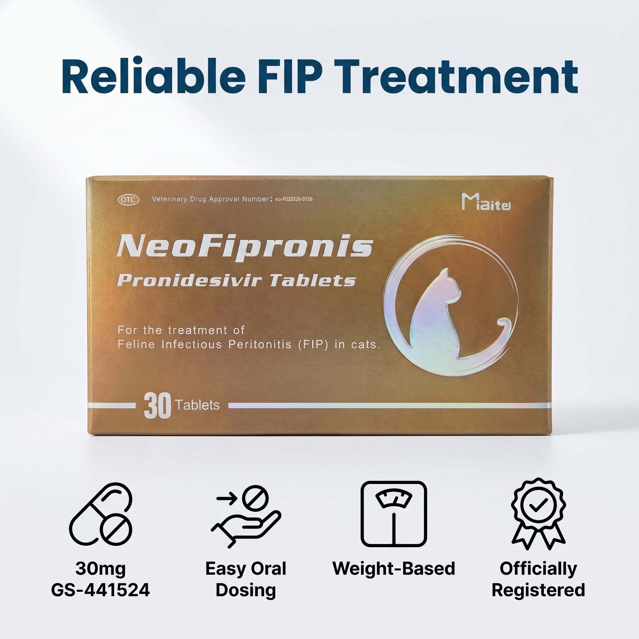
Currently, FIP remains a challenging disease to treat, with few effective options. Recent advances have introduced antiviral drugs like GS-441524 showing promise, but access and approval vary geographically. Treatment often aims to manage symptoms and reduce inflammation, including corticosteroids or immunosuppressants, which may temporarily improve ocular symptoms.
The prognosis depends heavily on the extent of systemic and neurological involvement. Cats with ocular FIP may experience temporary visual improvement with treatment, but the disease is typically progressive. Preventing vision loss remains difficult, emphasizing early detection and supportive care.
Preventive Measures and Owner Awareness
Since FIP results from a mutation of feline coronavirus, controlling environmental factors and managing high-risk populations can reduce incidence. Good hygiene, minimizing stress, and avoiding overcrowding decrease the risk of coronavirus mutations.
Owners should be vigilant for behavior changes, eye abnormalities, or neurological signs. Early veterinary consultation can improve management outcomes and quality of life, even in terminal cases.
Final Thoughts
While FIP more commonly affects internal organs, it has the potential to impact a cat’s vision through ocular and neurological involvement. Recognizing the signs early and seeking veterinary care are critical steps in managing affected cats. Understanding the disease’s complexity underscores the importance of preventive measures and ongoing research for better treatment options.
References
1. Pedersen, N. C. (2014). An overview of feline infectious peritonitis virus infection and disease. The Veterinary Journal, 200(1), 17-21.
2. Addie, D. D., et al. (2009). Feline coronavirus: Pathogenesis and clinical presentation. Veterinary Clinics of North America: Small Animal Practice, 39(4), 641-652.
3. Hartmann, K. (2005). Feline infectious peritonitis. The Veterinary Clinics of North America: Small Animal Practice, 35(1), 39-79.
4. Kipar, A., & Detmer, S. (2015). Feline coronavirus: Pathogenesis, diagnosis, and management of FIP. Veterinary Medicine and Science, 1(4), 243-255.
5. Pimentel, N., et al. (2020). Advances in Feline Infectious Peritonitis Treatment. Frontiers in Veterinary Science, 7, 570.