What Indicators Are Monitored During FIP Follow-Up Exams

Feline Infectious Peritonitis (FIP) remains one of the most challenging diseases in feline medicine due to its complex pathogenesis and variable clinical presentation. Although recent advancements have improved diagnostic accuracy and treatment options, ongoing monitoring remains essential for assessing disease progression and response to therapy. During follow-up examinations, veterinarians focus on a range of clinical and laboratory indicators to inform management decisions and optimize feline health.
Clinical Signs and Physical Examination
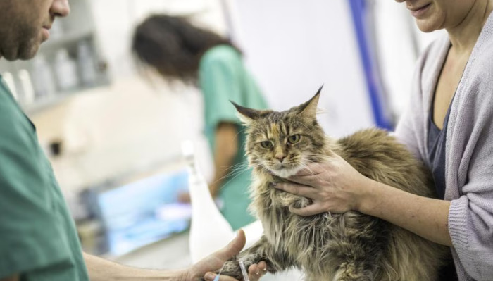
The initial step in FIP follow-up is a comprehensive physical examination. Clinicians observe for changes in weight, body condition, hydration status, and temperature. FIP often presents with signs like abdominal distension, weight loss, lethargy, or fever. Monitoring these parameters helps determine if the disease remains controlled or if there’s evidence of clinical progression.
Auscultation of the thorax and abdomen provides additional insights. For instance, a muffled heart or lungs may suggest effusion accumulation, while abdominal masses or distension could indicate ongoing inflammatory processes or fluid buildup. Gentle palpation can also assess the presence of lymphadenopathy or organomegaly, common in FIP-afflicted cats.
Serum and Biochemical Parameters
Blood tests are critical during follow-up. Complete Blood Count (CBC) often reveals anemia, lymphopenia, or neutrophilia. Anemia in FIP may be regenerative or non-regenerative, directly corresponding to the disease’s impact on marrow or chronic inflammatory state.
Serum chemistry panels are essential for evaluating organ function. Elevated liver enzymes such as ALT and ALP can indicate hepatic involvement or inflammation. Changes in serum albumin and globulin levels are particularly significant: decreased albumin combined with increased globulins (especially gamma globulins) are characteristic signs of ongoing immune response or inflammation, providing clues about the disease’s activity.
Monitoring for hypoproteinemia or hyperglobulinemia assists in assessing disease severity. Persistent or worsening biochemical abnormalities may suggest active disease or complications requiring intervention.
Imaging Studies
Diagnostic imaging modalities like ultrasound and radiography play pivotal roles. Serial ultrasounds help evaluate the presence and quantity of effusions in the abdomen or thorax, and detect abnormalities such as organ enlargement or masses.
Ultrasound can also assess the integrity of the kidneys, liver, and lymph nodes, providing visual confirmation of inflammatory or infiltrative processes. In cases of ocular or neurological FIP, other imaging techniques like MRI or CT can offer detailed evaluations.
Serial imaging during follow-up helps detect disease progression or response to therapy based on regressive or progressive changes in effusions and organ structures.
Cerebrospinal Fluid (CSF) and Effusion Analysis
For cats with neurological or ocular signs, sampling and analyzing CSF or effusions is vital. During follow-up, analysis focuses on detecting inflammatory changes, such as increased protein, cellularity, or specific cell types.
In FIP, CSF or effusion fluid often presents with elevated protein levels, a high non-degenerate neutrophil count, and increased globulin levels. Cytology, alongside molecular diagnostics like RT-PCR, can confirm ongoing infection.
Serial fluid analyses enable clinicians to evaluate if inflammatory parameters are decreasing, indicating a positive response, or worsening, which might suggest ongoing or refractory disease.
Serological and Molecular Testing
While antibody detection is useful for initial diagnosis, it has limited value in follow-up because of persistent antibody titers even after resolution. Conversely, quantitative PCR (qPCR) for FCoV RNA in tissues, blood, CSF, or effusions can quantitatively assess viral load dynamics over time.
Monitoring viral RNA levels during follow-up enables veterinarians to gauge disease activity and response to antiviral treatments. A decrease in viral load often correlates with clinical improvement, while persistent or increasing viral RNA may signal ongoing infection.
Immunological Markers and Cytokines
Emerging research suggests that profiling cytokines and immune markers can offer insights into the inflammatory environment within affected tissues. Elevated levels of pro-inflammatory cytokines like IL-6, IL-1β, and TNF-α are associated with active disease. Monitoring these markers might help assess immune response and guide immunomodulatory therapies.
Treatment Response Indicators
In cases where antiviral agents such as GS-441524 are used, monitoring their efficacy involves observing clinical improvement in conjunction with laboratory indicators. A sustained reduction in effusions, normalization of blood parameters, and decreasing viral RNA levels are positive signs of therapeutic success.
Conversely, lack of improvement or worsening parameters call for a reassessment of treatment strategy or supportive care adjustments. Regular updates on these indicators allow for timely modifications aimed at improving prognosis.
Conclusion
Monitoring during FIP follow-up exams involves a multifaceted approach combining clinical assessments, laboratory tests, imaging studies, and molecular diagnostics. Each indicator offers a piece of the puzzle, collectively enabling veterinarians to evaluate disease activity, response to therapy, and long-term prognosis. As research advances, the integration of emerging biomarkers and diagnostic techniques holds promise for even more precise disease management.
References
1. Pedersen, N. C. (2014). An overview of feline infectious peritonitis virus infection and immune responses. Veterinary Microbiology, 181(1-2), 41–49.
2. Addie, D. D., et al. (2016). Feline infectious peritonitis: ABCD guidelines on prevention and management. Journal of Feline Medicine and Surgery, 18(7), 575–583.
3. Hartmann, K. (2019). Feline infectious peritonitis. Veterinary Clinics of North America: Small Animal Practice, 49(4), 887–899.
4. Kipar, A., & Meli, M. L. (2014). Feline infectious peritonitis: still an enigma. Veterinary Pathology, 51(2), 505–526.
5. Felten, S., et al. (2017). Diagnostic challenges of feline infectious peritonitis. Veterinary Clinical Pathology, 46(4), 518–527.