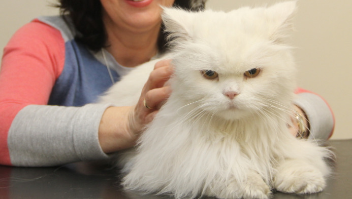
What Every Cat Owner Should Know About the FIP Diagnostic Process
Feline Infectious Peritonitis (FIP) has long been regarded as one of the most challenging and devastating diseases affecting domestic cats. While ongoing research and treatment advancements have begun to reshape the outlook for affected cats, the diagnostic process for FIP remains complex and often frustrating for pet owners and veterinarians alike. Understanding the nuances of this process is essential for anyone who cares for a cat, especially when symptoms emerge that might suggest FIP. This article dives deep into the details of FIP diagnosis, outlining the scientific background, clinical experience, and practical steps that guide veterinarians in their search for answers.
Understanding Feline Infectious Peritonitis (FIP)
At its core, FIP is an illness resulting from infection with a mutant form of feline coronavirus (FCoV). Most domestic cats worldwide carry feline coronavirus at some point without issues, but a small number will experience the transformation of this benign virus into the form responsible for FIP. The disease can manifest in a “wet” (effusive) form, where fluid accumulates in body cavities, and a “dry” (non-effusive) form, often involving organ inflammation with fewer obvious outward signs.
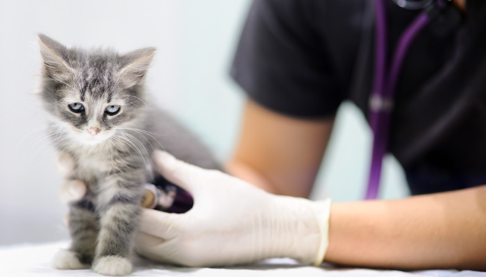
FIP is classically seen in young cats, but it can occur in any age group. Risk factors include living in multi-cat environments, genetic susceptibility, and immunocompromising conditions. The disease is always regarded as fatal without intervention, but new antiviral drugs have begun offering hope, provided an accurate diagnosis is achieved early.
Why Is FIP So Difficult to Diagnose?
FIP does not present with hallmark symptoms exclusive to its pathology. Instead, it often begins with vague signs: lethargy, poor appetite, weight loss, fever, or a swollen abdomen. Many other feline diseases cause similar problems. The gold standard for FIP diagnosis would be detection of the virus within affected tissues by immunohistochemistry—a process often only possible after death. Therefore, the challenge is to distinguish FIP from other illnesses using a combination of clinical suspicion, laboratory tests, and advanced diagnostics, rather than a single “yes or no” result.
The universal feline coronavirus complicates matters. Tests that detect feline coronavirus antibodies or RNA cannot differentiate between “normal” coronavirus infection and the mutated, disease-causing FIP form. This diagnostic ambiguity is a major source of confusion and distress for cat owners.
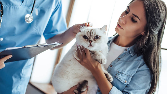
The Role of Clinical History and Examination
Veterinarians begin the diagnostic process by gathering a comprehensive history. Key details include the cat’s age, breed, environment, recent exposures, and the progression of clinical signs. Breed predisposition is notable; for example, Bengals, Birman, and Ragdoll cats may be overrepresented in FIP cases.
The physical exam can reveal clues, especially in classic “wet” FIP: fluid accumulation in the abdomen (ascites) or chest (pleural effusion), jaundice, or palpable organ masses. “Dry” FIP may present only with vague neurological signs, ocular manifestations like inflammation inside the eye, or lymph node enlargement.
Laboratory Diagnosis: The Puzzle Pieces
Bloodwork and biochemical analysis provide essential information. Almost all cats with FIP will show abnormalities, but not every abnormality is diagnostic. Hallmarks include:
Hyperglobulinemia: a raised total globulin level in the blood, due to immune activation
Low albumin:globulin ratio (usually <0.8), which increases FIP suspicion
Mild to moderate anemia (low red blood cells)
Lymphopenia (low lymphocyte counts)
High bilirubin indicating liver dysfunction
Routine laboratory studies alone cannot confirm FIP, but they help prioritize additional tests. A complete blood count and chemistry panel are always considered the foundation.
Effusion Analysis: Fluid Clues
For cats with wet FIP, fluid analysis from the abdomen or chest is particularly informative. Veterinarians will assess the color, viscosity, protein content, and cell count:
Effusions are usually straw-colored, clear, with high protein (>3.5 g/dL), and relatively low cell counts.
Rivalta test: a simple procedure carried out to distinguish FIP-related effusion from other causes. A positive Rivalta test increases suspicion but is not definitive.
Effusion samples may also be sent for PCR, cytology, and specific protein evaluation. The absence of bacteria and neoplastic cells in the fluid further tips the scale toward an FIP diagnosis.
Serological Testing: The Coronavirus Challenge
Most FIP cases occur in cats that test positive for feline coronavirus exposure. Veterinarians will run ELISA or immunofluorescent antibody tests to detect antibodies. High antibody titers may support FIP suspicion in symptomatic cats, but many healthy cats show similar results. Recent advances in PCR technology allow for detection of coronavirus RNA in blood or effusion. Still, the presence of RNA is not unique to FIP and cannot distinguish the mutant form.
Emerging tests focus on the FIP-specific nucleoprotein and unique genetic markers, but these remain limited in availability and validation.
Imaging: Ultrasound and Beyond
Ultrasound is a useful tool, especially for detecting fluid in body cavities, masses, or enlargement of organs like the spleen, liver, and kidneys. In dry FIP, imaging assists in identifying subtle organ changes, lymphadenopathy, and thickening of intestinal walls. Sometimes computed tomography (CT) or magnetic resonance imaging (MRI) become necessary, particularly for neurological FIP.
Radiographs are not considered highly informative, as FIP rarely produces specific radiographic signs. However, chest radiographs may confirm pleural effusion and exclusion of heart disease.
Tissue Sampling and Biopsy: The Gold Standard
The most definitive method for confirming FIP is finding the virus within tissue lesions using immunohistochemistry or immunofluorescence. Tissue samples are typically obtained from affected organs during surgery or, rarely, post-mortem. This gold standard is impractical in most living cats due to invasiveness, risk, and expense.
Recent advances highlight the use of fine-needle aspirates from lymph nodes, abdominal masses, or organs, but the diagnostic yield is lower than full tissue biopsies.
In most cases, diagnosis rests on “strong suspicion” based on clinical signs, laboratory data, imaging, and exclusion of other diseases rather than direct proof.
Differential Diagnosis: Ruling Out Other Diseases
FIP overlaps with several other feline diseases. Excluding alternative causes is crucial:
Lymphoma and other neoplasias can mimic the abdominal masses and effusions
Bacterial, fungal, or protozoal infections may produce similar fluid and systemic signs
Immune-mediated diseases cause chronic fever and organ inflammation
Heart disease can result in fluid accumulation (ascites or pleural effusion)
Veterinarians use a stepwise elimination approach, often ruling out other diseases before arriving at FIP.
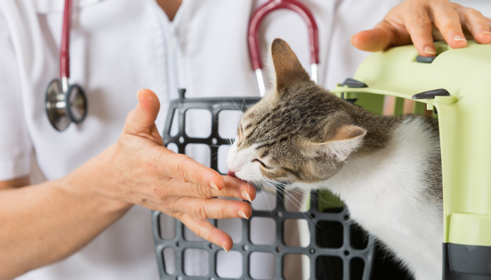
Considerations for Multi-Cat Households
FIP is not considered highly contagious, but its parent virus (feline coronavirus) is easily spread in multi-cat environments through shared litter boxes and grooming. Diagnosing FIP in one cat prompts consideration for the health status of others. Testing healthy cats for feline coronavirus is not routinely recommended, as positivity does not predict FIP.
Cat owners in shelters, catteries, and foster homes must remain vigilant for signs. Strategies include reducing stress, practicing good hygiene, and minimizing overcrowding.
Advancements in Molecular Testing
Genetic and molecular research have led to more sophisticated tests for FIP. These include:
Reverse Transcriptase PCR (RT-PCR) for viral RNA in tissues, fluids, or blood
Immunohistochemistry for viral protein within tissue samples
Some PCR assays target specific mutations associated with FIP, differentiating them from wild-type feline coronavirus. Availability is improving, but not all tests are widely accessible or standardized.
The Implications of a Diagnosis
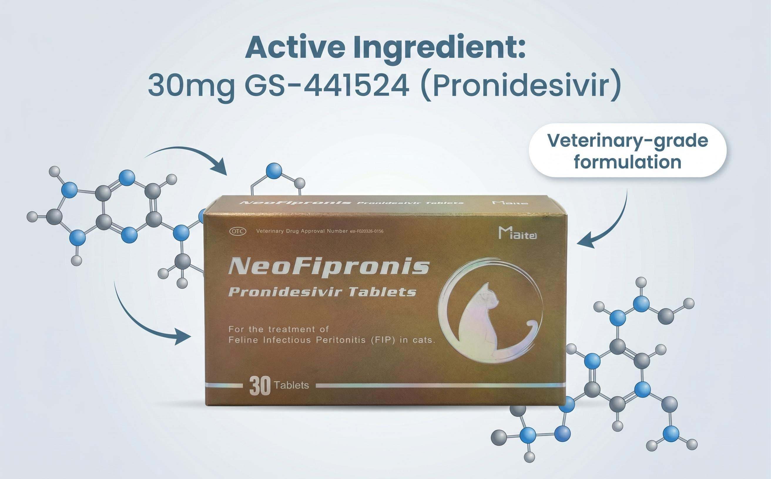
Confirmation of FIP has historically carried a grave prognosis. Until recently, there were no reliable treatments. However, investigational antiviral drugs, especially GS-441524 and its analogs, have changed the landscape for many cats—offering remission in a substantial proportion with early intervention.
Diagnosis also impacts multi-cat households. Pet owners must consider environmental decontamination, prudent quarantine, and psychological support for themselves and affected cats.
Communication Between Owner and Veterinarian
The diagnostic process for FIP can be deeply emotional. Owners often face uncertainty and distress due to the stepwise, often inconclusive nature of testing. Veterinarians walk a fine line, balancing clinical suspicion with objective data, always aiming to deliver up-to-date and compassionate guidance.
Support groups, online resources, and referral to feline infectious disease specialists may improve understanding and decision-making during this challenging process.
Living With the Uncertainty
A diagnosis of FIP is rarely black and white. Many cats are classified as “suspected FIP” based on exclusion of other diseases and medical imaging, without tissue confirmation. When laboratory data and clinical signs strongly point toward FIP, the risk of misdiagnosis decreases, especially in young cats with classic effusion and compatible bloodwork.
Owners must prepare for ambiguity and collaborate closely with their veterinarian to weigh risks, benefits, and response to antiviral therapy.
The Future of FIP Diagnosis
Owing to rapid advances in molecular techniques, genetic sequencing, and global research collaboration, the pathway to more precise and less invasive diagnostics continues to improve. Efforts are underway to standardize high-specificity genetic tests capable of distinguishing FIP mutations from benign feline coronavirus strains.
Until then, diagnosing FIP remains an art informed by science—a careful blend of clinical observation, laboratory analysis, imaging, and, in select cases, molecular testing.
References
1. Pedersen NC. A review of feline infectious peritonitis virus infection: 1963–2008. J Feline Med Surg. 2009;11(4):225-258.
2. Hartmann K. Feline infectious peritonitis. Vet Clin North Am Small Anim Pract. 2005;35(1):39–79.
3. Tasker S. Diagnosis of feline infectious peritonitis: update on evidence supporting laboratory testing and future developments. Vet J. 2018;237:54-60.
4. Addie DD, Graham E, Krzemińska M, et al. Feline coronavirus, FCoV, and FIP: The facts, the myths, and the mysteries. Vet Rec. 2020;186(3):94-98.
5. Chang HW, Egberink HF, Halpin R, et al. Spike protein fusion peptide and feline coronavirus type I/II serotyping. J Gen Virol. 2012;93(8):1627-1638.
6. Felten S, Hartmann K. Diagnosis of feline infectious peritonitis: a review of the current literature. Viruses. 2019;11(11):1060.
7. Kipar A, Meli ML. Feline infectious peritonitis: Still an enigma? Vet Pathol. 2014;51(2):505-526.
8. Dempsey SM, Ewing PJ. A review of the pathogenesis, clinical diagnosis, and management of feline infectious peritonitis. J Vet Intern Med. 2011;25(4):791-803.
9. Ishida T, Shibanai A, Tanaka S, et al. Use of recombinant feline interferon and glucocorticoids in the treatment of feline infectious peritonitis. J Feline Med Surg. 2004;6(2):107-109.
10. Veterinary Manual, Merck & Co., Inc. Feline Infectious Peritonitis. Available: https://www.merckvetmanual.com
11. American Association of Feline Practitioners. FIP Guidelines. Available: https://catvets.com/guidelines/practice-guidelines/feline-infectious-peritonitis
12. University of California, Davis. Feline Infectious Peritonitis (FIP) Research. Available: https://www.vetmed.ucdavis.edu
13. Norsworthy GD, Crystal MA, Grace S. FIP in the era of new drugs: Advances in feline medicine. J Feline Med Surg. 2022;24(9):e695-703.