Different Types of FIP
Feline Infectious Peritonitis (FIP) remains one of the most challenging and complex diseases affecting domestic cats. It is caused by a mutated form of the feline coronavirus (FCoV), which can lead to various clinical presentations. Recognizing the different types of FIP is crucial for accurate diagnosis, treatment, and management. The disease manifests primarily in two major forms: effusive (wet) FIP and non-effusive (dry) FIP, each exhibiting distinct pathophysiological features and clinical signs.
Effusive (Wet) FIP
Wet FIP is characterized by the accumulation of serous fluid within body cavities, predominantly in the abdomen or thorax. This form is often more acute and dramatic, involving a robust inflammatory response. The pathogenesis involves immune complex formation that leads to vasculitis, increased vascular permeability, and subsequent fluid leakage. Cats with wet FIP typically present with signs such as abdominal distension, difficulty breathing, fever unresponsive to antibiotics, and weight loss. Laboratory findings often reveal hyperproteinemia, high globulin levels, and moderate anemia. The rapid progression of fluid accumulation distinguishes wet FIP from other conditions with similar signs.
Non-effusive (Dry) FIP
The dry form of FIP presents without significant fluid accumulation. Instead, granulomatous inflammatory lesions develop in various organs, such as the kidneys, liver, eyes, and central nervous system. This variant is more insidious, with symptoms varying based on the organs involved. Common signs include weight loss, persistent fever, ocular abnormalities like uveitis, neurological deficits, and lethargy. Histopathologically, dry FIP exhibits granulomas rich in macrophages, lymphocytes, and plasma cells. Laboratory tests may show elevated globulin levels and specific antibody titers, but fluid analysis usually shows little to no effusion, making diagnosis more challenging.
Mixed (Intermediate) FIP
While classification primarily revolves around wet and dry forms, an intermediate presentation exists. Cats exhibiting both effusive and granulomatous features display mixed FIP. These cases may show fluid accumulation alongside granulomatous lesions in various tissues. Such presentations underscore the disease's heterogeneity and complicate differential diagnosis. Approximating the prevalence of mixed FIP is difficult, but veterinary practitioners should remain vigilant for overlapping signs.
Variations Based on Organ Involvement
FIP's clinical diversity extends beyond the wet-dry classification. Depending on the organs affected, the disease may be further subdivided:
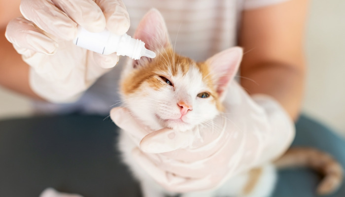
Ocular FIP: Presents primarily with uveitis, retinal detachment, and other eye abnormalities. Often seen in dry FIP.
Neurological FIP: Characterized by meningoencephalitis, ataxia, cranial nerve deficits, and seizures. It can occur in both wet and dry forms but is more common in dry FIP.
Peritonitis and Hepatic FIP: Involving the abdominal organs, leading to ascites, hepatomegaly, and abdominal pain.
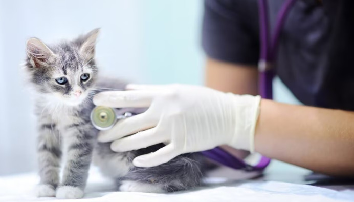
Pathological Spectrum and Diagnosis
The different FIP types demonstrate a spectrum where immune response and viral mutation influence clinical presentation. The effusive form is associated with a more humoral immune response, leading to vasculitis and fluid leakage, whereas the dry form involves a more cell-mediated response, resulting in granuloma formation. Diagnostic differentiation relies on clinical signs, laboratory testing—such as total protein levels, globulin ratios, and specific antibody titers—and histopathology. Imaging techniques like ultrasound can reveal fluid accumulation or granulomatous lesions, aiding in identifying the FIP type.
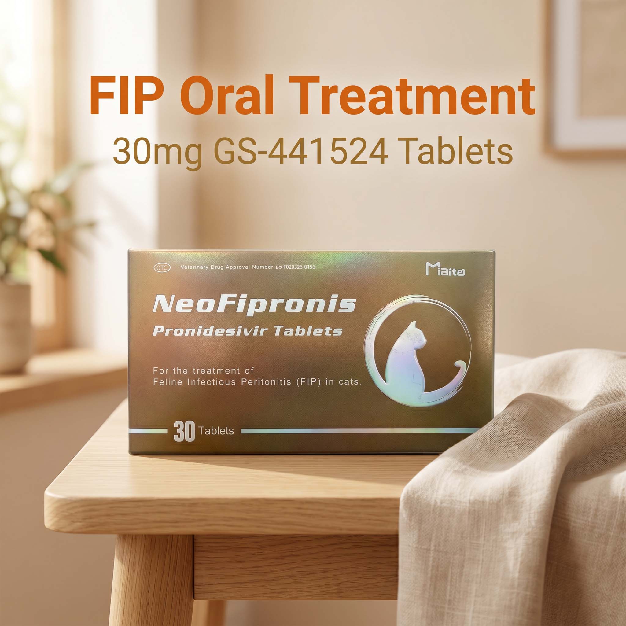
Implications for Treatment and Prognosis
While antiviral therapies and immunomodulators have shown promise, the prognosis remains guarded, particularly for wet FIP due to its rapid progression. Recognizing the FIP type can influence treatment decisions; for example, anti-inflammatory therapy may benefit dry FIP more than wet FIP. Additionally, understanding the disease's form aids in providing owners with realistic expectations and palliative options when necessary.
Unique Challenges in Veterinary Practice
The heterogeneity of FIP complicates the establishment of a one-size-fits-all diagnostic or treatment protocol. The overlap of clinical signs among different forms often leads to misdiagnosis or delayed intervention. Emerging diagnostic tools, including PCR assays and advanced imaging, are paving the way for more precise identification of FIP variants. Continued research into the molecular mechanisms differentiating wet and dry FIP is vital for developing targeted therapies.
References
1. Pedersen, N. C. (2014). An Overview of Feline Infectious Peritonitis Virus Pathogenesis and Diagnosis. Veterinary Microbiology, 171(3-4), 221-227.
2. Addie, D. D., & Jarrett, O. (2006). Feline Infectious Peritonitis. In Feline Medicine and Therapeutics (pp. 403-418).
3. Kipar, A., & Meli, M. L. (2014). Feline Infectious Peritonitis: Still an Enigma? Journal of Feline Medicine and Surgery, 16(6), 528-533.
4. Hartmann, K. (2014). Feline Infectious Peritonitis. Veterinary Clinics of North America: Small Animal Practice, 44(4), 791-804.