What Does the Color of Abdominal Fluid Indicate in FIP
Feline Infectious Peritonitis (FIP) is one of the most challenging diseases affecting cats. It results from an aberrant immune response to feline coronavirus infection. Although coronavirus infection in cats is common and typically benign, a small percentage of cats develop FIP, which is almost invariably fatal without prompt and appropriate intervention. Among its hallmark features, abnormal fluid accumulation in the abdomen or chest—known as effusion—is a dire sign and a clue for diagnosis. The color, consistency, and other physical characteristics of this fluid can reveal crucial clinical information. Understanding what the color of abdominal fluid means in FIP is essential for veterinarians and pet owners navigating this complex condition.
Abdominal Effusion in FIP: Definition and Pathogenesis
FIP develops when mutated feline coronavirus escapes containment by the immune system and triggers a destructive inflammatory response. The effusive (wet) form of FIP is characterized by leakage of protein-rich fluid into body cavities, most commonly the abdomen. This results from damage to the blood vessels caused by immune complexes, which increases vascular permeability, allowing fluid components to leak out.
Effusion in FIP is typically a straw-yellow to golden color, but it can sometimes present with other colors, such as clear, cloudy, or even bloody. The color arises from the composition of the fluid, the underlying inflammatory process, and possible secondary complications.
Yellow or Straw-Golden Fluid: The Classic FIP Indicator
The most recognizable color of abdominal effusion in FIP is a bright yellow or straw-golden hue. This color is so characteristic that it is frequently mentioned in textbooks and diagnostic guidelines. The main reason for this color is the high protein content – notably globulins – giving the fluid an almost “sticky” appearance, sometimes creating a web-like effect when handled.
Several factors contribute to this yellow color:
Bilirubin: This pigment, resulting from the breakdown of hemoglobin, accumulates when there is extensive inflammation and cellular destruction. Although not always present in pathological amounts, bilirubin can contribute a yellowish tinge.
Protein Content: Total protein is usually greater than 3.5 g/dL. Albumin and globulins suspended in the fluid reflect light in a way that produces a golden color.
Inflammation: The inflammatory response triggered by FIP leads to exudation of plasma proteins, cellular debris, and inflammatory cells, which blend to give the effusion its distinctive hue.
Clear or Light-Colored Effusion: Unusual, but Possible
Not all abdominal effusions in FIP are the vivid yellow that textbooks describe. Early in the disease process or in less severe cases, the fluid may be more transparent or pale. In some cases, especially with rapid accumulation, the color may be less intense than expected.
Other causes for clear effusions include:
Dilution by Intravenous Fluids: In hospitalized cats receiving IV fluids, the concentration of proteins and pigments may fall, rendering the fluid less colored.
Early Stage FIP: At the onset of effusion formation, the protein and cellular content may not yet reach levels needed to tint the fluid yellow.
Mixed Forms: Some cats have both effusive and non-effusive FIP; in these cases, the fluid may not fit the classical description.
Cloudy or Turbid Fluid: Sign of Secondary Complications
Cloudiness usually suggests the presence of increased white blood cells, bacteria, or lipid particles. While the classic FIP effusion is more clear and viscous, secondary bacterial infections, concurrent pancreatitis, or trauma may all cloud the fluid.
Key causes for turbidity:
White Blood Cells: Intense inflammation can promote the migration of leukocytes, making the fluid cloudy.
Bacteria: FIP fluid is typically sterile, but secondary infection from intestinal perforation or translocation may result in cloudy, foul-smelling fluid.
Chyle: Milky fluid can result from lymphatic leakage—though this is rare in FIP.
Red or Blood-Tinged Fluid: Hemorrhage or Trauma
Blood within the abdominal fluid carries a different implication. While FIP alone generally does not cause frank hemorrhage, severe vasculitis may rarely allow blood components to escape into the abdominal cavity. Procedures such as abdominal taps (paracentesis) can also cause iatrogenic bleeding.
Potential sources of bloody fluid include:
Trauma: Cat fights, falls, or other injuries.
Severe Vasculitis: Extensive damage to blood vessels from unchecked inflammation.
Concurrent Disease: Neoplasia, coagulopathies, or other conditions affecting blood vessels.
It is important to distinguish from other diseases that cause bloody abdominal effusion, such as hemangiosarcoma, rodenticide toxicity, or ruptured organs.
Green or Brown Fluid: Infection, Bile or GI Contents
Rarely, the abdominal fluid may be green, brown, or otherwise abnormal in color. These findings almost always suggest an additional, more acute pathology:
Bile Peritonitis: Leakage of bile from the gallbladder or biliary tract causes green fluid and signals a surgical emergency.
Intestinal Rupture: Leakage of gastrointestinal contents is brown and malodorous and requires urgent care.
Necrotizing Infections: Overwhelming bacterial infection can alter fluid color dramatically.
Such presentations are not typical of FIP alone and warrant thorough evaluation.
How Color Guides Diagnosis
Veterinarians rely heavily on the appearance and laboratory analysis of abdominal fluid to distinguish FIP from other conditions. The typical straw-yellow, viscous fluid with high protein and cellular content argues strongly for FIP when accompanied by appropriate clinical signs (fever, weight loss, abdominal distension, and organomegaly). However, fluid color alone cannot confirm FIP; laboratory tests such as measurement of the albumin-to-globulin ratio, Rivalta test, cytology, PCR for FCoV RNA, and antibody titers are essential.
When the fluid is not classic in appearance, veterinarians look for additional signs:
Cloudy or purulent fluid raises suspicion for bacterial peritonitis.
Green or brown fluid suggests GI rupture or bile leakage.
Bloody fluid mandates investigation for trauma, neoplasia, or severe vasculitis.
Because abdominal fluid color can overlap among multiple diseases, veterinarians always interpret it alongside the cat’s full clinical context and diagnostic workup.
Laboratory Analysis: Beyond Visual Inspection
Beyond simple color assessment, a full laboratory analysis offers vital data:
Total Protein Measurement: Effusions associated with FIP almost always exceed 3.5 g/dL.
Cellularity: The fluid contains a mixed population of neutrophils, macrophages, and sometimes lymphocytes.
Albumin/Globulin Ratio: FIP fluid often shows a low ratio (< 0.8).
Rivalta Test: This bedside test can help differentiate FIP effusion from others.
Cytological Examination: Smears may reveal phagocytic cells and abundant proteinaceous material.
PCR and Immunohistochemistry: These test for the presence of coronavirus within effusion cells for definitive diagnosis.
Lab results help resolve ambiguous findings and differentiate FIP from heart failure, liver disease, neoplasia, or bacterial peritonitis.
Differential Diagnosis: Other Causes of Abdominal Fluid
It is important to compare FIP effusion with fluid from other disease processes:
Congestive Heart Failure: Usually clear and low protein content.
Liver Disease: May be yellow, but typically less viscous than FIP fluid.
Neoplasia: Can produce all fluid types, sometimes bloody.
Septic Peritonitis: Oftentimes cloudy, foul-smelling, and may contain bacteria.
Hypoproteinemia: Usually clear, watery fluid.
Veterinarians always maintain a broad differential when assessing abdominal fluid in cats.
Clinical Approach: Integrating Fluid Color into the FIP Diagnosis
When confronted with a cat presenting with abdominal distension, fever, and malaise, a veterinarian will typically:
1. Perform imaging (ultrasound or radiographs) to confirm the presence and volume of fluid.
2. Collect fluid via paracentesis, evaluating its color, viscosity, and odor.
3. Submit the sample for laboratory analysis: protein, cell count, cytologic examination, Rivalta test, and PCR or antibody testing for FCoV.
4. Integrate fluid findings with clinical history, physical exam, and other laboratory data.
The spectrum of fluid color forms part of this comprehensive diagnostic process. In the context of FIP, a bright yellow, sticky, protein-rich effusion argues immediately for FIP, though it requires confirmation.
Fluid Color and Prognosis in FIP
While fluid color helps guide diagnosis, it does not usually indicate prognosis. The severity of clinical signs—such as anemia, jaundice, rapidly increasing effusion volume, or evidence of organ dysfunction—are more relevant to outcome.
In some cases, chronic, slow-accumulating fluid may appear less yellow over time as protein content lags. Rapid accumulation resulting in tense abdominal distension may signal a more aggressive disease course.
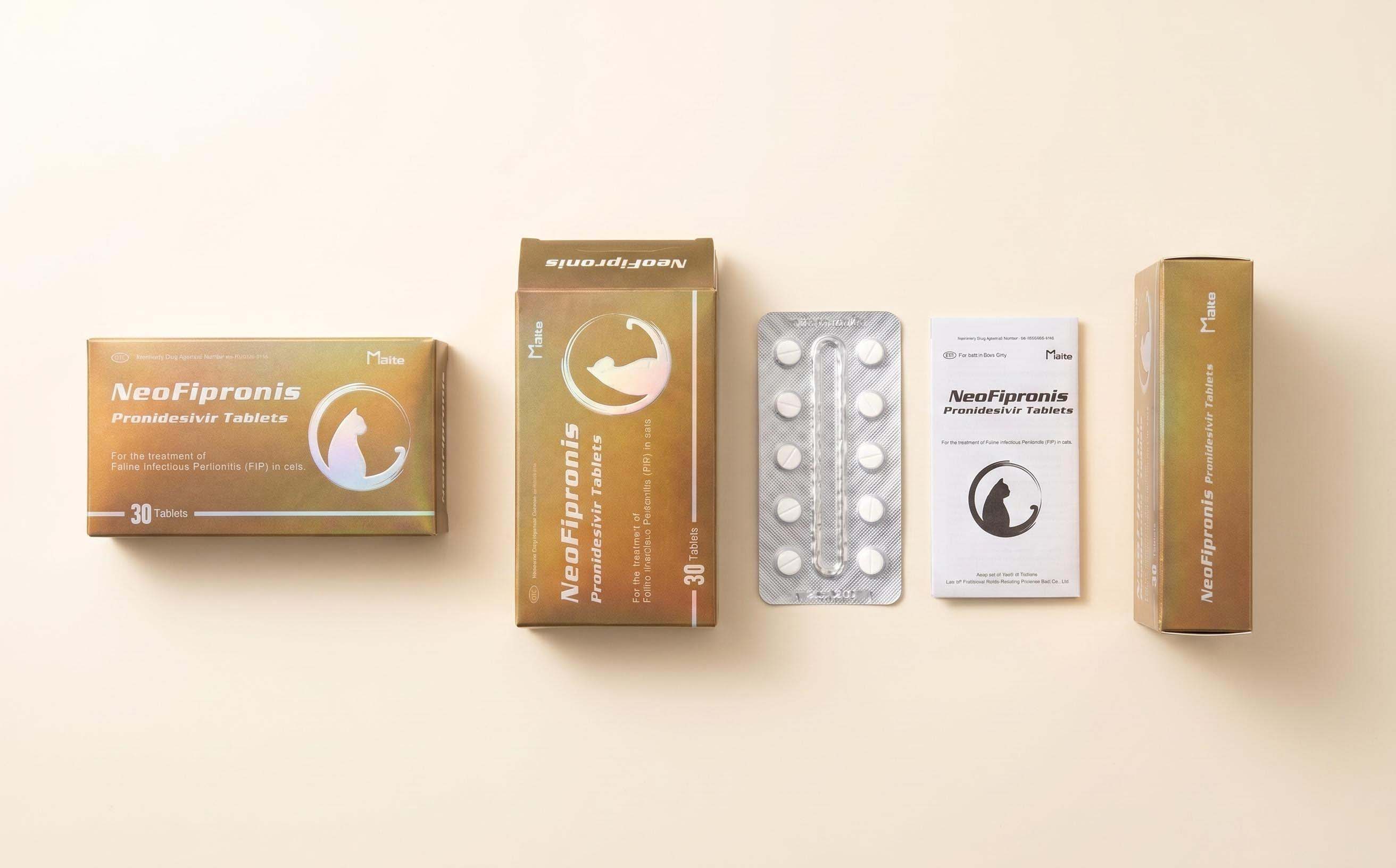
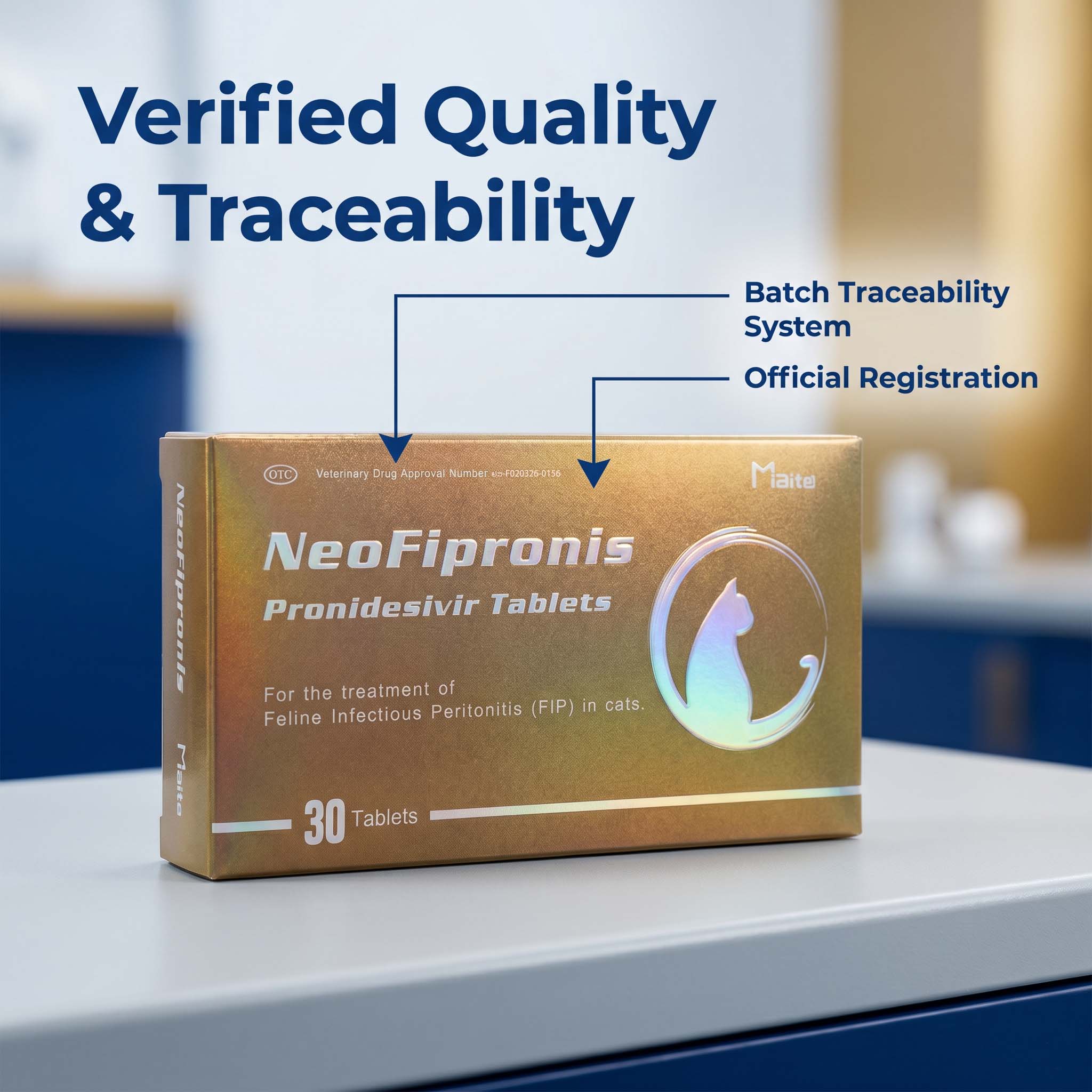
Treatment Advances and Monitoring
Recent years have seen rapid advances in FIP management, with antiviral drugs such as GS-441524 and its derivatives offering the possibility of remission. Effusion color can sometimes be used to monitor response to treatment; successful therapy typically leads to resolution of effusion, and fluid color normalization often precedes clinical improvement.
Nevertheless, repeated paracentesis should be avoided unless fluid is compromising breathing or comfort, as it provides only temporary relief.
Client Communication: What Pet Owners Should Know
When discussing abdominal fluid with cat owners, veterinarians must clarify that fluid color is only part of the puzzle. While a yellow fluid is suggestive of FIP, it is not enough for a definitive diagnosis—especially since other diseases can produce effusions of similar color. Owners should be advised about the need for confirmatory testing, and the urgent nature of FIP diagnosis and treatment.
Any sudden change in fluid color, or worsening of clinical signs (lethargy, vomiting, breathing difficulty) demands immediate veterinary attention.
Implications for Future Research
Rapid diagnostic tools for FIP effusion analysis remain an active area of research. New markers, point-of-care tests, and methods for coronavirus detection in effusion cells are being developed. Improved understanding of effusion color and composition may lead to faster, more accurate diagnosis, and better outcomes for affected cats.
References
1. Hartmann, K. (2005). Feline infectious peritonitis. Veterinary Clinics: Small Animal Practice, 35(1), 39-79.
2. Pedersen, N. C. (2014). An update on feline infectious peritonitis: diagnostics and therapeutics. Veterinary Journal, 201(2), 133-141.
3. Dempsey, S. M., & Ewing, P. J. (2011). Feline infectious peritonitis: diagnostic dilemmas. Compendium: Continuing Education for Veterinarians, 33(8), E1–E8.
4. Tasker, S. (2018). Diagnosis of feline infectious peritonitis: Update on evidence supporting laboratory tests. Journal of Feline Medicine and Surgery, 20(3), 228-243.
5. Kipar, A., & Meli, M. L. (2014). Feline infectious peritonitis: still an enigma?. Veterinary Pathology, 51(2), 505-526.
6. Addie, D. D., & Jarrett, O. (1992). Feline coronavirus antibodies in cats. Veterinary Record, 131(10), 225-226.
7. Fishman, E. J., & Patterson, M. M. (2003). The Rivalta test for feline infectious peritonitis. Journal of the American Veterinary Medical Association, 223(2), 174-179.
8. Felten, S., & Hartmann, K. (2019). Diagnosis of feline infectious peritonitis: a review of the current literature. Viruses, 11(11), 1068.