Viral Infection Mechanism of FIP

Feline Infectious Peritonitis (FIP) stands as one of the most enigmatic and devastating infectious diseases affecting domestic cats worldwide. Triggered by certain mutations in feline coronavirus (FCoV), FIP presents a unique challenge to veterinarians and cat owners. This article explores the molecular and cellular mechanisms underlying the viral infection process of FIP, focusing on viral entry, immune system interaction, pathogenesis, and current research directions. Coverage includes the transition from benign enteric coronavirus to pathogenic FIP virus, immune evasion tactics, and tissue tropism. This information benefits the veterinary community, researchers, and cat owners seeking to understand the intricacies of FIP infection.
Feline Coronavirus: The Parent Virus
Feline coronavirus (FCoV) is a common virus found in cat populations globally. Most infections result in mild or asymptomatic enteric disease. The two recognized types of FCoV are FECV (Feline Enteric Coronavirus) and FIPV (FIP Virus). The development of FIP occurs when the relatively harmless FECV undergoes mutations, granting new abilities that enable systemic spread and severe disease. FIPV evolves within the host either due to random mutations or through recombination events, often during persistent infections.
Mechanisms of Mutation: From FECV to FIPV
Research highlights that one critical step in FIP pathogenesis is the mutation in the viral spike (S) protein gene. This alteration allows the virus to infect monocytes and macrophages, escaping initial containment in the gut. The proposed mutation occurs in the S gene’s furin cleavage site, enabling a change in cell tropism. This change is pivotal, as it marks the virus’s ability to disseminate throughout the cat’s body and initiate life-threatening systemic infection.
Other mutations in non-structural proteins may also play a role, affecting the virus’s replication efficiency and evasion of host immune responses. The diversity of FCoV strains and their variable mutation rates make FIP emergence unpredictable and difficult to prevent in multi-cat environments.
Viral Entry and Replication
Once mutated, FIPV has enhanced capability to bind to feline cellular receptors. Entry involves attachment to complementary receptors on monocytes and macrophages, followed by fusion of the viral envelope with the cell membrane. Post-entry, viral RNA is uncoated and hijacks the cell’s translational machinery, generating viral proteins and new genomic RNA. This process depends largely on the ability of viral nonstructural proteins to manipulate host translation and block immune detection.
Replication sites, known as double-membrane vesicles, serve as a protected area for viral RNA synthesis. These organelles shield the virus from intracellular defenses, establishing persistent infection. Newly formed virions exit by exocytosis or budding, ready to infect additional host cells.
Immune Evasion Strategies
Central to FIPV’s pathogenicity is its exceptional arsenal of immune evasion tools. Infected monocytes and macrophages become Trojan horses, disseminating the virus.
1. Antibody-Dependent Enhancement (ADE): Rather than neutralizing the virus, some antibodies generated against FCoV facilitate infection. ADE occurs when antibodies bind the virus without inactivating it, promoting uptake into macrophages through Fc receptor-mediated endocytosis.
2. Inhibition of Antigen Presentation: FIPV can block efficient antigen presentation, minimizing activation of effective T cell responses.
3. Interferon Antagonism: Nonstructural proteins block interferon signaling cascades, dampening antiviral defenses and allowing viral replication to proceed unchecked.
4. Induction of Apoptosis in Lymphoid Tissues: The virus directly induces apoptosis in lymphoid cells, contributing to immunosuppression and making the host more vulnerable to secondary infections.
Pathogenesis and Granulomatous Inflammation
FIP development hinges on the host’s immune response. While some cats clear the infection, others develop aggressive granulomatous inflammation. The immune system’s fight against infected monocytes leads to the formation of pyogranulomas—collections of neutrophils, macrophages, and lymphocytes surrounding sites of viral replication. Aberrant vasculitis, characterized by immune-mediated damage to blood vessels, is a hallmark of FIP pathology.
There are two clinical manifestations of FIP:
1. Effusive (Wet) Form: Marked by accumulation of protein-rich effusions in body cavities (abdomen, thorax), a consequence of severe vasculitis and plasma leakage. Cats often present with abdominal distension, respiratory distress, and rapid progression.
2. Non-Effusive (Dry) Form: Characterized by granuloma formation in various organs (kidneys, liver, CNS, eyes) without significant effusion. Symptoms vary based on the affected organ system—neurological signs, ocular changes, or organ dysfunction may predominate.
The outcome is determined by the balance between destructive humoral responses and protective cell-mediated immunity. Poor cellular immune response leads to progressive FIP, while robust T-cell activity may contain or eliminate infection.
Transmission and Host Factors
FIP is not considered contagious in its mutated, systemic form. Transmission occurs primarily via fecal-oral spread of FECV, with the pathogenic mutation arising within individual cats. High-density living conditions, young age, genetic susceptibility, and chronic stress have all been identified as risk factors for FIP development. Breeds such as British Shorthair, Birman, and Bengal cats show increased predisposition, likely due to underlying immunogenetic differences.
Key Cellular Interactions
Central to FIPV spread is the interaction between the virus and monocytes/macrophages. The S protein’s affinity for targeted host cell receptors is critical. Following internalization, these immune cells travel in the bloodstream and lymphatics, bringing the virus to diverse tissues. The inability of macrophages to efficiently kill the mutated virus allows for persistent infection and widespread granuloma formation.
Endothelial cells lining blood vessels are collateral victims of the intense immune reaction, leading to increased permeability and effusion formation. The cyclical nature of viral infection, immune attack, and subsequent tissue damage results in the classical peritonitis seen in FIP.
Diagnostic Challenges
Diagnosing FIP remains a formidable task in veterinary medicine. No single test offers perfect specificity and sensitivity due to the close genetic relationship between FECV and FIPV. RT-PCR may detect viral RNA but cannot always distinguish between enteric and FIP-associated strains. Diagnosis often relies on a combination of clinical signs, cytological analysis of effusions, elevated protein levels, and imaging findings. Immunohistochemistry and PCR targeting specific mutations in S gene are emerging tools for definitive diagnosis.
Therapeutic Approaches and Research Frontiers
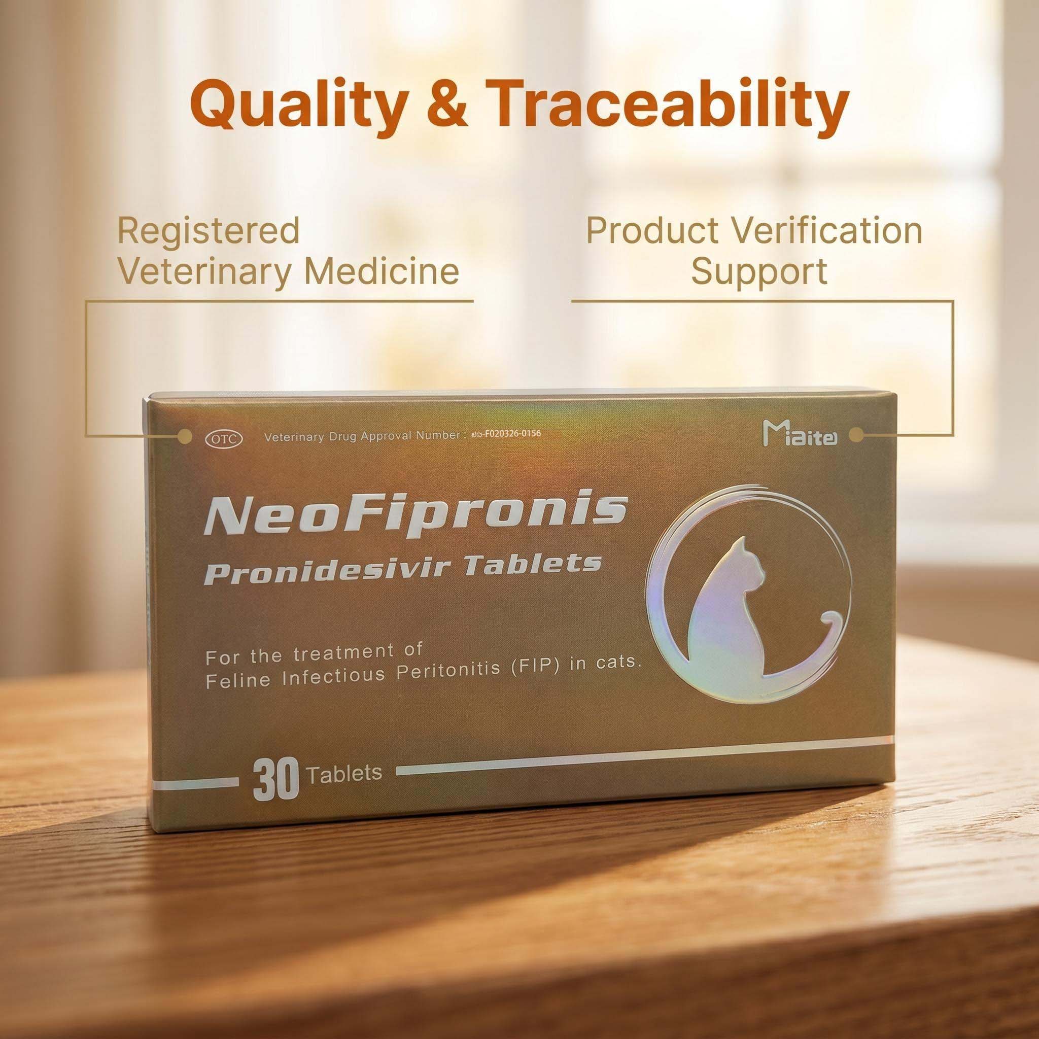
For decades, FIP lacked effective treatment. Recent advances have revolutionized the outlook for affected cats. Protease inhibitors (GS-441524, a nucleoside analogue related to remdesivir) disrupt viral replication and have demonstrated promising results. Antiviral therapy prolongs survival and can lead to remission in many cases.
Immunomodulatory therapies target the excessive inflammatory response, sometimes with modest success. Research continues to elucidate the genetic basis of susceptibility and resistance, with the hope of informing future vaccine development. Prevention strategies focus on limiting exposure in high-risk environments, maintaining low population density, and prompt removal of fecal material.
Future Directions in FIP Mechanism Studies
The molecular biology of FIPV mutation, entry, tropism, and immune evasion remains an area of active investigation. Novel sequencing technologies enable detailed characterization of viral populations and mutation patterns. Understanding host genetic factors that predispose to FIP will be critical in developing targeted interventions. Vaccines have thus far met with limited success due to the complex immune paradox posed by ADE.
Strategies for future research include:
1. Mapping S gene mutations and their impact on cell tropism.
2. Elucidating host genetic variations in cytokine response and cellular immunity.
3. Identifying new antiviral targets in FIPV’s replicative machinery.
4. Determining biomarkers for early and accurate FIP diagnosis.
Conclusion
Breakthroughs in understanding FIP’s viral infection mechanisms have transformed the outlook for affected cats but much remains to be discovered. Central features include virus mutation, monocyte/macrophage infection, immune evasion, and granulomatous inflammation. As therapies and diagnostics advance, the need for continued research and public education grows. Addressing the multifaceted biology of FIP and its viral origins will require cooperation across research disciplines, veterinary professionals, and the pet-owning community.
References
1. Pedersen NC. A review of feline infectious peritonitis virus infection: 1963–2008. Journal of Feline Medicine and Surgery. 2009;11(4):225-258.
2. Chang HW, Egberink HF, Halpin R, et al. Use of naturally occurring mutants to analyze the functions of the feline coronavirus replication complex. Journal of Virology. 2012;86(18):8973-8981.
3. Kipar A, Meli ML. Feline infectious peritonitis: still an enigma? Veterinary Pathology. 2014;51(2):505-526.
4. Tekes G, Thiel HJ. Feline coronavirus: pathogenesis of feline infectious peritonitis. Viruses. 2016;8(6):pii:E127.
5. Felten S, Hartmann K. Diagnosis of feline infectious peritonitis: A review of the current literature. Viruses. 2019;11(11):1068.
6. Pearson M, LaVoy A, Evans S, et al. Intracellular retention of mutant spike protein causes endoplasmic reticulum stress in feline infectious peritonitis. Journal of General Virology. 2019;100(2):192-206.
7. Murphy BG, Perron M, Murakami E, et al. The nucleoside analog GS-441524 strongly inhibits feline infectious peritonitis (FIP) virus in tissue culture and experimental cat infection studies. Veterinary Microbiology. 2018;219:226-233.
8. Vennema H, Poland A, Foley J, et al. Feline infectious peritonitis viruses arise by mutation from endemic feline enteric coronaviruses. Virology. 1998;243(1):150-157.
9. Pedersen NC. Serologic and virologic findings in cats with natural and experimental feline infectious peritonitis. American Journal of Veterinary Research. 1976;37(6):815-820.
10. Dewerchin HL, Cornelissen E, Nauwynck HJ. Replication of feline coronaviruses in peripheral blood monocytes. Archives of Virology. 2005;150(2):2483-2500.