Is It Possible to 100% Confirm FIP
Introduction: Understanding Feline Infectious Peritonitis (FIP)
Feline Infectious Peritonitis (FIP) persists as one of the most daunting and perplexing diseases in feline medicine. Caused by a mutation of feline coronavirus (FCoV), FIP presents an intricate diagnostic challenge. The virus is incredibly common, with surveys indicating up to 40% or more cats worldwide may carry FCoV, particularly in multi-cat environments. Yet FIP itself is considered a rare yet devastating consequence—a product of immune-mediated reaction to mutated FCoV. Its varied clinical manifestations and similarity to other diseases frequently lead both owners and veterinarians to confront one central question: Is it ever possible to 100% confirm FIP in a living cat?
The Science Behind FIP and Its Mutation
FIP is the result of a specific mutation within the ubiquitous feline coronavirus. While the majority of infected cats experience mild and self-limiting enteric symptoms, a small subset undergoes a viral mutation that enables the virus to infect macrophages, pivotal immune cells. This switch from benign GI infection to a systemic, often fatal disease alters everything: the virus disseminates, triggering immune-mediated vasculitis and multi-organ inflammation.
There are two main forms of FIP: effusive (wet) and non-effusive (dry). Each displays characteristic immune responses, but both can present overlapping symptoms, making FIP a “disease of mimicry.” This mutability and similarity in clinical signs with other common feline illnesses create the core problem in FIP diagnostics.
Clinical Signs and Their Diagnostic Value
Initial signs of FIP are nonspecific and include lethargy, loss of appetite, and fever unresponsive to antibiotics. As the disease progresses, effusive or ‘wet’ FIP leads to fluid accumulation in body cavities, often the abdomen and/or thorax. Dry FIP, in contrast, creates granulomatous lesions in organs like the kidney, liver, or CNS, complicating the clinical picture further.
Despite these hallmark features, individual symptoms are not pathognomonic. Similar presentations are found with other diseases such as lymphoma, bacterial peritonitis, toxoplasmosis, and some metabolic conditions. This overlap leads clinicians to seek more definitive diagnostics instead of relying on symptoms alone.
Laboratory and Imaging Diagnostics: Valuable but Imperfect
Laboratory abnormalities in FIP are often suggestive but rarely definitive. Hematology may show anemia, lymphopenia, or neutrophilia; serum biochemistry might demonstrate elevated total protein, increased globulin, and decreased albumin with an altered albumin-to-globulin ratio. These patterns are commonly but not exclusively seen in FIP.
Analysis of effusions offers useful clues, as FIP fluid is typically straw-colored, viscous, with high protein content (>35g/L) and low cellularity. Rivalta’s test, though simple, is not absolutely conclusive—false positives and false negatives do occur.
Abdominal ultrasonography or thoracic imaging may reveal fluid accumulation or organ changes, yet these findings are not specific to FIP. MRI or CT may aid in investigating neurological or ocular FIP presentations but do not confirm the underlying cause.
Molecular Diagnostics: PCR and Immunohistochemistry
Modern diagnostics rely heavily on molecular techniques. Reverse transcriptase PCR (RT-PCR) can detect FCoV RNA in blood, tissue, or effusions. However, PCR is limited by issues of sensitivity and specificity: a positive PCR for FCoV indicates infection but not necessarily FIP, as many healthy carriers are PCR-positive. Quantitative PCR (qPCR) can help by measuring viral loads, but even this cannot single-handedly differentiate between enteric FCoV and mutated FIP viruses.
Immunohistochemistry (IHC), using antibodies to detect FCoV antigen within tissue macrophages, is currently regarded as the gold standard for FIP diagnosis. Detection in diseased tissue is highly suggestive of FIP, especially in context of clinical and histological findings. However, IHC requires invasive biopsy or post-mortem tissue, often unfeasible or unethical in living patients.
Antibody Testing and Serology: Diagnostic Challenges
Serological tests measure anti-FCoV antibodies in blood or fluids. High titers suggest exposure but do not reflect FIP mutation or progression. The ubiquity of FCoV in cat populations retards any useful specificity; cats without disease often have high titers, while cats with FIP can occasionally show low or undetectable levels, especially in terminal stages when immune collapse occurs.
Genetic Sequencing and Advances in FIP Diagnosis
Recent advances aim to distinguish the specific FIP mutation via genetic sequencing. Studies have identified mutations in the spike protein (S gene) unique to FIP-pathogenic virus strains, offering theoretical capability for precise detection. Yet, even with advanced sequencing, overlap persists, as not all S gene mutations correlate reliably with clinical disease. Sensitivity and specificity remain imperfect; wide-scale screening is cost-prohibitive and inaccessible to most veterinary practitioners.
Histopathology: The Gold Standard Revisited
Histopathological exam, ideally coupled with IHC, remains the definitive diagnostic approach. Seeing characteristic pyogranulomatous inflammation, vasculitis, and FCoV antigen within tissue macrophages offers the highest confidence. Nonetheless, the need for invasive sampling (laparotomy, organ biopsy) makes this method impractical for living cats. Often, diagnosis is only confirmed post-mortem, creating the jarring reality that living FIP diagnosis is always a matter of best evidence rather than absolute certainty.
Clinical Diagnosis and the Problem of Probability
Most FIP diagnoses in practice rest upon a probabilistic framework. Veterinarians use a “weight-of-evidence” approach, factoring together history, clinical signs, laboratory findings, imaging, response to therapy, and molecular diagnostics. Algorithms such as the Cornell FIP Diagnostic Tree recommend scoring systems and criteria-based checklists. Ultimately, even in cases where everything points to FIP, some margin of error exists—other diseases can rarely mimic FIP with extraordinary accuracy.
Novel Therapies and the Diagnostic Imperative
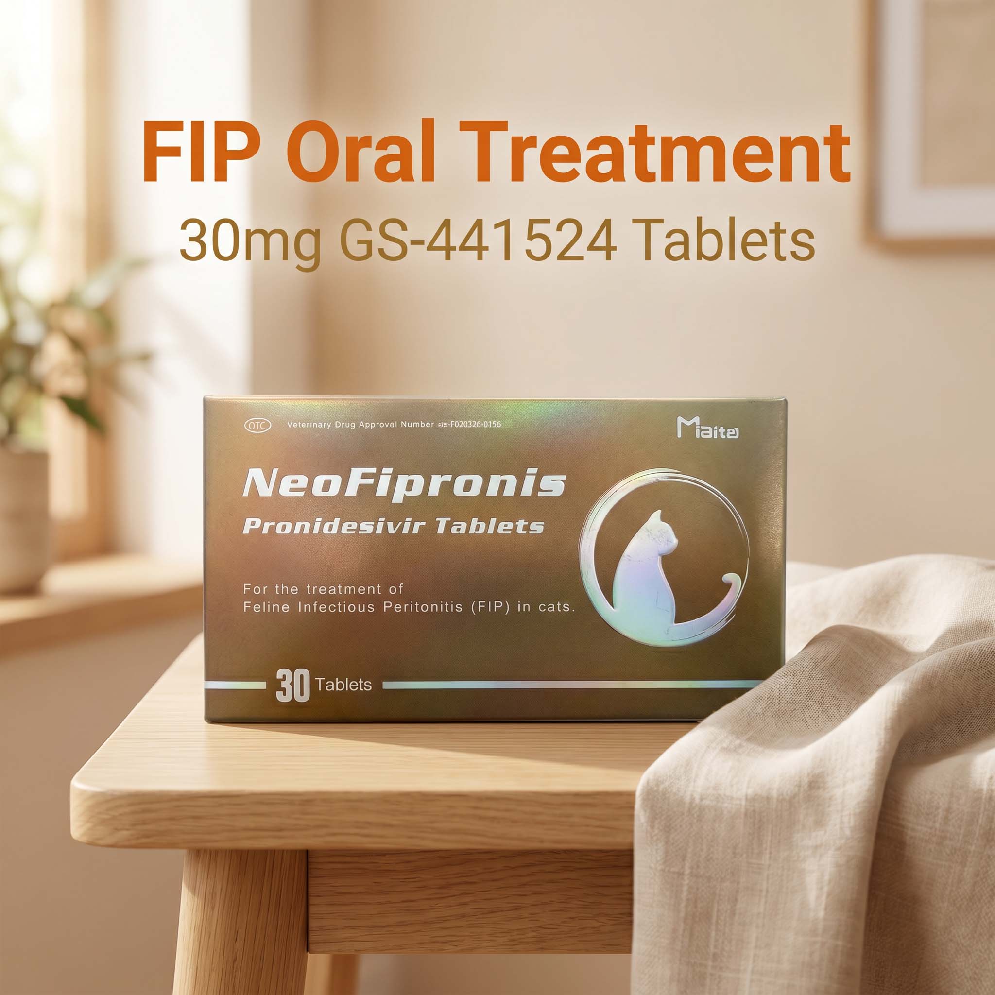
The recent emergence of antiviral therapies, such as GS-441524 and remdesivir, have offered remarkable hope, with survival rates of eighty to ninety percent when treatment is initiated early. Ironically, however, these advances intensify diagnostic demands: unnecessary treatment is costly and exposes animals to drug risks, while missed FIP diagnoses can be fatal.
Thus, practitioners are pressed to make diagnoses with maximal confidence, but must grapple with imperfect tests and the persistent possibility of error. Some clinics may opt for empirical treatment based on strong suspicion; others delay, seeking additional confirmatory evidence.
Current Recommendations for Clinicians and Cat Owners
Guiding principles include gathering comprehensive history (crowded environment, young age, recent stressors), paying close attention to suggestive signs (persistent fever, weight loss, abdominal/thoracic fluid), using multi-modal diagnostic strategies (laboratory, imaging, molecular/serology, cytology), and consulting FIP diagnostic guidelines or specialist advice.
Given the present limitations, communication is key. Owners should understand that a “100% diagnosis” of FIP is rarely achievable in real time. Decisions should be made collaboratively, balancing diagnostic evidence, disease likelihood, and the risks and benefits of potential interventions.
Ethical Considerations in Diagnostic Certainty
The inability to confirm FIP with absolute certainty has ethical consequences. Serial testing, invasive procedures, or unnecessary euthanasia should be weighed against the feasibility of empirical treatment and supportive care. Most veterinary professionals advocate for the “best clinical diagnosis” model, prioritizing the welfare of the cat within a framework of responsible medicine.
Future Directions in FIP Diagnostics
Research continues into new diagnostic strategies, such as non-invasive tissue imaging, targeted genetic tests, and improved serological markers. The hope is to one day offer real-time, point-of-care, definitive FIP diagnosis that is practical, affordable, and non-invasive. Until then, FIP remains the “great pretender”—demanding cautious optimism and collaborative trust among veterinarians and cat owners alike.
References
1. Pedersen, N. C. (2009). A review of feline infectious peritonitis virus infection: 1963–2008. Journal of Feline Medicine and Surgery, 11(4), 225-258.
2. Kipar, A., & Meli, M. L. (2014). Feline infectious peritonitis: Still an enigma? Veterinary Pathology, 51(2), 505-526.
3. Addie, D. D., et al. (2020). Feline infectious peritonitis: ABCD guidelines on prevention and management. Journal of Feline Medicine and Surgery, 22(7), 631-638.
4. Felten, S., & Hartmann, K. (2019). Diagnosis of feline infectious peritonitis: A review of available methods and their limitations. Veterinary Journal, 245, 29-37.
5. Tasker, S. (2018). Feline coronavirus infections: Update on diagnostics and therapeutics. Veterinary Clinics: Small Animal Practice, 48(1), 1-23.
6. Stranieri, A., et al. (2018). Diagnostic performance of FCoV RT-PCR in blood and effusions for FIP diagnosis. Journal of Feline Medicine and Surgery, 20(4), 378-385.
7. Barker, E. N., et al. (2017). Feline coronavirus with and without spike gene mutations detected by real-time reverse transcriptase PCR in cats with and without feline infectious peritonitis. Journal of General Virology, 98(6), 1675-1682.
8. Dempsey, S. M., & Ewing, P. J. (2011). Infectious diseases of the gastrointestinal tract. Clinical Techniques in Small Animal Practice, 16(3), 202-211.
9. Carrau, T., et al. (2021). Real-world efficacy of GS-441524-based therapy for FIP. Journal of Feline Medicine and Surgery, 23(6), 667-673.
10. Boyd, D. F., et al. (2022). Advances in molecular diagnostics and treatment of feline infectious peritonitis. Veterinary Clinics: Small Animal Practice, 52(1), 111-128.
11. American Association of Feline Practitioners (AAFP). (2022). Feline Infectious Peritonitis Diagnosis and Management Guidelines.
12. Kornreich, B. G. (2019). Clinical decisions in suspected feline infectious peritonitis: Weighing probabilities and evidence. Veterinary Clinics: Small Animal Practice, 49(1), 37-55.