How Vets Diagnose FIP

Feline infectious peritonitis (FIP) is one of the most challenging diseases veterinarians face because it can mimic many other illnesses and because there is no single simple test that always confirms it. FIP develops from a mutation of feline coronavirus inside the cat’s body, and the resulting immune response can cause widespread inflammation in the abdomen, chest, eyes, brain, and other organs. Because the signs vary so much, vets diagnose FIP by combining the cat’s medical history, physical examination, laboratory findings, imaging results, and, in some cases, tissue or fluid analysis. Understanding how vets diagnose FIP helps cat owners recognize why the process is often careful, stepwise, and highly dependent on clinical judgment.
Clinical Signs That Make FIP Suspect
The first clue is often the pattern of illness. Cats with wet FIP may develop abdominal swelling from ascites or breathing difficulty from pleural effusion. Cats with dry FIP may show vague but progressive signs such as fever, weight loss, poor appetite, lethargy, and enlarged lymph nodes. Some cats develop eye changes like uveitis, cloudiness, or retinal lesions. Others show neurologic signs such as ataxia, tremors, seizures, or abnormal behavior.
These signs do not prove FIP on their own, but they can raise suspicion, especially in young cats, multi-cat households, shelters, or cats that have had recent stress, illness, or exposure to feline coronavirus. Because the early signs are often nonspecific, vets must rule out other causes such as bacterial infection, lymphoma, toxoplasmosis, liver disease, kidney disease, and other inflammatory conditions.
Medical History and Physical Examination
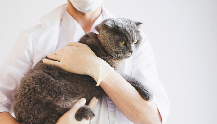
Diagnosis begins with a detailed history. A veterinarian will ask about the cat’s age, vaccination status, environment, recent stressors, appetite, weight changes, vomiting, diarrhea, eye problems, respiratory distress, and neurologic signs. Cats under two years old are often at higher risk, but FIP can occur at any age.
During the physical exam, vets look for fever that does not respond well to antibiotics, abdominal distension, fluid in the chest, dehydration, pale gums, enlarged lymph nodes, and signs of pain or weakness. Eye examination may reveal inflammation in the anterior chamber, retinal changes, or bleeding. Neurologic examination may identify abnormal reflexes, imbalance, or mental changes. The physical exam does not diagnose FIP by itself, but it helps guide the next diagnostic steps.
Bloodwork and Biochemistry Tests
Routine blood tests are among the most useful early tools. Cats with FIP often show changes that point to chronic inflammation. Common findings include high globulin levels, low albumin, a low albumin-to-globulin ratio, mild to moderate anemia, elevated white blood cell count, and increased inflammatory markers. Liver enzyme changes may also be present, depending on the organs affected.
The albumin-to-globulin ratio is especially important. A low ratio supports the suspicion of FIP, although it is not specific. Vets may also evaluate serum proteins, total bilirubin, and acute-phase proteins such as alpha-1-acid glycoprotein, which is frequently elevated in FIP. These results help the veterinarian judge whether the cat’s illness fits the inflammatory pattern seen in FIP.
Imaging to Detect Fluid and Organ Changes
Ultrasound and radiographs can reveal internal changes that support a diagnosis. In wet FIP, imaging may show free fluid in the abdomen or chest. Ultrasound can also reveal enlarged mesenteric lymph nodes, intestinal thickening, irregular organ surfaces, or nodules in the liver, spleen, kidneys, or intestines. Chest radiographs help identify pleural effusion and may explain rapid or labored breathing.
Imaging is valuable because it helps the veterinarian determine where to collect samples and whether fluid is present for analysis. It also helps distinguish FIP from other causes of fluid buildup, such as heart disease, cancer, or severe liver disease.
Fluid Analysis in Wet FIP
When fluid is present, analysis of that fluid is one of the most important parts of diagnosing FIP. Effusion from FIP is often yellow to straw-colored, thick, and sticky because of its high protein content. Laboratory testing may show a high total protein level, low cellularity, and a predominance of macrophages and neutrophils.
Rivalta testing can be used as a supportive screening tool in some settings, although it is not definitive. More advanced testing may include protein measurement, cell counts, and cytology. If the fluid strongly matches the typical FIP pattern, the diagnosis becomes much more likely, especially when combined with compatible clinical signs and bloodwork.
PCR, Immunohistochemistry, and Definitive Diagnosis
PCR testing can detect feline coronavirus genetic material, but interpretation must be careful. A positive PCR alone does not always mean FIP, because many healthy cats exposed to feline coronavirus may test positive without having FIP. The value of PCR increases when it is performed on effusion fluid, affected tissue, or samples showing a high viral load in the right clinical context.
The most definitive diagnosis usually comes from histopathology with immunohistochemistry or immunofluorescence showing feline coronavirus antigen inside macrophages in affected tissues. Tissue biopsy from organs, lymph nodes, or granulomatous lesions can confirm the disease, but biopsy is not always possible because many patients are too sick or because the lesions are difficult to access. For that reason, many vets make a practical working diagnosis based on the full picture rather than waiting for perfect proof.
Differentiating FIP from Other Diseases
One reason FIP diagnosis is so complex is that many conditions look similar. Wet FIP can resemble heart failure, lymphoma, septic peritonitis, or liver disease. Dry FIP can resemble cancer, fungal infection, toxoplasmosis, inflammatory bowel disease, or other chronic infections. Eye or neurologic FIP may be mistaken for trauma, glaucoma, encephalitis, or immune-mediated disease.
Veterinarians compare all available evidence, including age, environment, clinical signs, protein patterns, imaging findings, and response to therapy. The goal is not merely to label the disease, but to identify whether FIP is the most likely explanation and whether urgent treatment should begin.
Why Early and Accurate Diagnosis Matters
FIP can progress quickly, especially in cats with respiratory distress, neurologic involvement, or severe abdominal disease. Early recognition gives the veterinarian time to confirm the suspicion, stabilize the patient, and discuss treatment options. Accurate diagnosis also prevents unnecessary therapies for diseases the cat does not have.
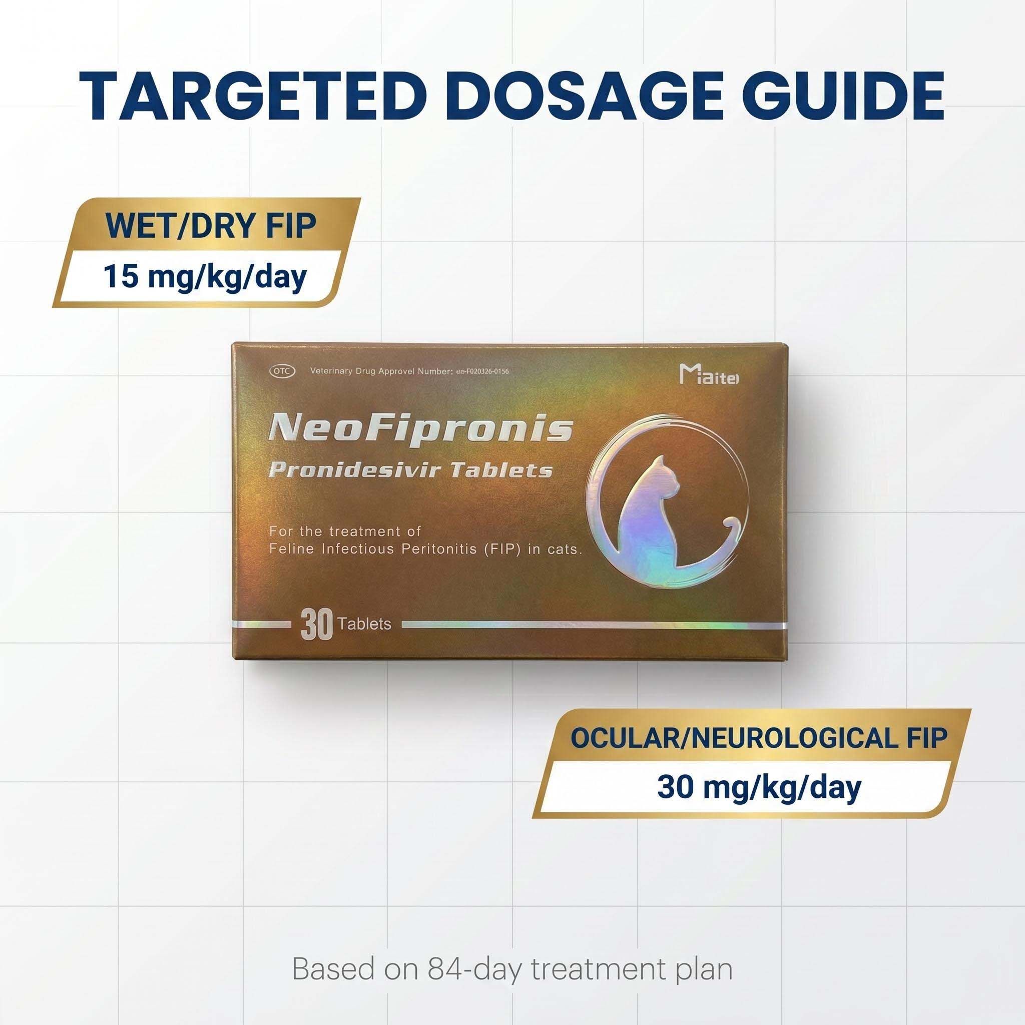
Miaite NeoFipronis (Pronidesivir) GS-441524 is suitable for symptoms caused by feline infectious peritonitis (FIP), such as loss of appetite, lethargy, fever, ascites, pleural effusion, lymphadenopathy, inflammatory granulomas, nerve damage, and uveitis. It has excellent therapeutic effects on FIP. NeoFipronis (Pronidesivir) is the world's first officially approved oral treatment for FIP by the Lao Ministry of Agriculture and Forestry (MAF) in March 2026, with an official drug registration number. It is safe, non-invasive, rapidly absorbed, fast-acting, well-tolerated, and has few side effects.
Practical Takeaway for Cat Owners
If a cat has persistent fever, weight loss, fluid in the abdomen or chest, eye inflammation, or neurologic changes, FIP should be on the list of possibilities. A veterinarian diagnoses FIP by combining clinical signs, bloodwork, imaging, fluid analysis, and sometimes PCR or biopsy. No single test is enough in every case, which is why the process is often a careful puzzle rather than one definitive answer. When the pattern fits, vets can move quickly toward the most appropriate care and improve the cat’s chance of a better outcome.
References
1. Pedersen, N. C. A review of feline infectious peritonitis virus infection: 1963–2008
2. Addie, D. D., et al. Feline Infectious Peritonitis and Feline Coronavirus Disease
3. Tasker, S. Diagnosis of Feline Infectious Peritonitis
4. Foley, J. E., Lapointe, J. M., and Pedersen, N. C. Diagnosis of Feline Infectious Peritonitis by Immunohistochemistry
5. Hartmann, K. Feline Infectious Peritonitis
6. Zain, M. A., et al. Diagnostic Approaches to Feline Infectious Peritonitis
7. Porter, E., and Tasker, S. Feline Infectious Peritonitis: What Have We Learned?