What Do High Inflammatory Markers Suggest in FIP

Introduction and Overview of FIP
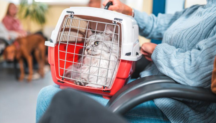
Feline Infectious Peritonitis (FIP) is a complex and often fatal disease that affects domestic and wild cats worldwide. Stemming from a mutation of feline coronavirus (FCoV), FIP manifests primarily in two forms: effusive (wet) and non-effusive (dry), both commonly associated with intense inflammation and immune dysregulation. Early diagnosis remains challenging due to overlapping symptoms with other feline illnesses, making laboratory diagnostics crucial. Inflammatory markers—both routine and advanced—play a critical role in understanding the pathogenesis, guiding clinicians in differentiation, disease monitoring, and prognostic evaluation.
Understanding Inflammatory Markers in Veterinary Medicine
Inflammatory markers are biomolecules produced or upregulated in response to infection, trauma, or autoimmunity. In cats, standard inflammatory markers include serum proteins such as alpha-1-acid glycoprotein (AGP), C-reactive protein (CRP), serum amyloid A (SAA), and globulins. Additional cellular markers associated with the hematological profile, such as leukocyte counts, and acute-phase cytokines like interleukin-6 (IL-6), also provide vital clues to underlying disease processes.
Routine veterinary panels may report increased total protein, globulin, and decreased albumin concentrations, alongside altered complete blood count (CBC) parameters in FIP cases. Reference ranges and interpretation may vary by laboratory, species, and individual patient characteristics. Elevated inflammatory markers specifically prompt practitioners to investigate systemic inflammation, guiding the differential diagnosis toward infectious etiologies such as FIP when other symptoms align.
The Role of Inflammatory Markers in the Pathogenesis of FIP
FIP occurs when feline coronavirus mutates within the host, enabling the virus to evade the immune system and infect macrophages. The immune system responds aggressively, sparking a sustained release of inflammatory mediators. These molecules damage blood vessels and cause pyogranulomatous lesions in organs. High concentrations of inflammatory proteins reflect the body's attempt to contain viral spread, but ironically, this overzealous response drives disease progression.
Key inflammatory markers in FIP include hyperglobulinemia (especially increased gamma globulins), elevated AGP, and increased SAA. Persistent high values not only indicate an active inflammatory process but also help distinguish FIP from other feline diseases such as neoplasia or bacterial peritonitis. In effusive FIP, hallmark findings include high protein exudate in the peritoneal or pleural cavities, mirroring the systemic inflammatory state.
Clinical Lab Findings in Cats With High Inflammatory Markers
Cats with FIP almost always exhibit an abnormal hematologic and biochemical profile. A CBC often reveals mature neutrophilia and lymphopenia, indicative of chronic inflammation driven by cytokine overproduction. Hyperglobulinemia, reflected as increased total protein with concurrent hypoalbuminemia, is a classic feature, raising the Albumin:Globulin (A:G) ratio red flag—often below 0.6 in FIP cases.
Alpha-1-acid glycoprotein is among the most sensitive acute-phase proteins in cats and is typically markedly elevated with FIP, especially during active disease. SAA and CRP also rise but are less specific, as they may be present in other infectious or inflammatory conditions. Thus, a panel approach, combining multiple markers with clinical and imaging data, increases diagnostic accuracy.
Interpreting High Inflammatory Markers: What Do They Suggest?
A marked increase in inflammatory markers prompts suspicion of systemic inflammation but is not exclusive to FIP. In the context of clinical presentation—such as fever, weight loss, abdominal distension, uveitis, or neurological symptoms—high inflammatory markers strongly suggest FIP, especially after ruling out common causes like bacterial infections, lymphoma, and other viral pathogens.
Interpretation should be multifactorial:
Diagnosis: While no single marker confirms FIP, a constellation of abnormalities (persistent high globulins, elevated AGP, lymphopenia, increased SAA) supports FIP diagnosis when coupled with clinical findings.
Disease Severity and Progression: Extremely elevated acute-phase proteins, especially AGP above 1500 µg/mL, often correlate with disease severity and a worse prognosis.
Therapy Monitoring: Successful antiviral therapy or immunomodulation should reduce inflammatory markers over time, signaling disease control or remission. Persistent elevation or renewed increase may warn of disease recurrence or progression.
Differentiating FIP From Other Conditions Using Inflammatory Markers
Many feline conditions, including bacterial peritonitis, pyelonephritis, neoplasia, immune-mediated diseases, and other viral infections, can cause elevated inflammatory markers. However, FIP typically produces a characteristic pattern: a disproportionately high globulin concentration with suppressed albumin, significant AGP elevation, and supportive clinical signs.
AGP and gamma globulins are most discriminative, while SAA and CRP, though sensitive, are less specific for FIP. Polymerase chain reaction (PCR) and immunohistochemistry for FCoV RNA or antigen in tissue samples, or effusions, remain diagnostic gold standards but inflammatory marker profiles help triage, select cases for advanced testing, and prioritize urgent intervention.
The Prognostic Value of Inflammatory Markers in FIP
Inflammatory markers offer valuable prognostic clues. Cats with extremely high AGP or persistent, non-resolving elevations of globulins and SAA tend to have a poorer prognosis. The Albumin:Globulin ratio proves useful; lower ratios indicate more severe disease and limited survival.
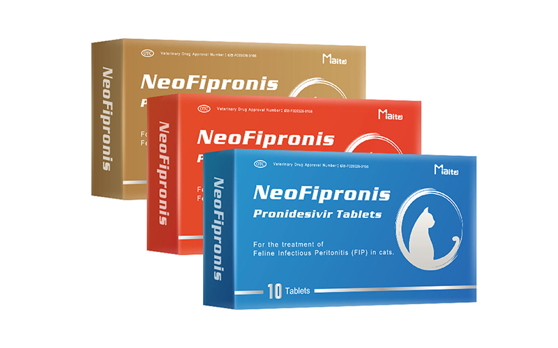
Emerging therapies for FIP—most notably GS-441524 and related antivirals—have shifted the historical paradigm from universally fatal to potentially treatable. Serial monitoring of inflammatory markers enables veterinarians to evaluate response to treatment, predict relapses, and adjust therapy as needed for optimal outcomes.
Emerging Research: Cytokines, Biomarkers, and Future Directions
Recent studies investigate novel inflammatory biomarkers such as interleukins (IL-1, IL-6), tumor necrosis factor-alpha (TNF-α), and other cytokine profiles in FIP-affected cats. These molecules help clarify the disease’s immunopathology, offering future diagnostic and therapeutic avenues.
Machine learning and artificial intelligence now analyze vast datasets of biochemical, imaging, and clinical parameters, improving predictive accuracy for FIP. This technology enables more precise risk stratification and monitoring, ultimately benefiting patient outcomes and advancing feline medicine.
Veterinary Practice: Practical Implications for Diagnosing and Managing FIP
Recognizing the significance of high inflammatory markers in cats with suggestive clinical signs is integral to veterinary practice. It allows for timely diagnosis, appropriate case selection for advanced testing, and informed client communication regarding prognosis and treatment options. As research continues, incorporation of novel biomarker assays promises even greater diagnostic clarity and better feline welfare.
References
1. Pedersen, N. C. (2009). A review of feline infectious peritonitis virus infection: 1963–2008. Journal of Feline Medicine and Surgery, 11(4), 225-258.
2. Dempsey, S. M., & Ewing, P. J. (2011). Feline infectious peritonitis: Diagnostic dilemmas and therapeutic options. Veterinary Medicine: Research and Reports, 2, 47-58.
3. Stranieri, A., Palmerini, D., Ceccherini, G., et al. (2018). Alpha-1 acid glycoprotein, feline coronavirus, and clinical outcome in feline infectious peritonitis. Journal of Feline Medicine and Surgery, 20(7), 653-661.
4. Hartmann, K. (2005). Feline infectious peritonitis. Veterinary Clinics of North America: Small Animal Practice, 35(1), 39-79.
5. Felten, S., & Hartmann, K. (2019). Diagnosis of feline infectious peritonitis: A review of the current literature. Viruses, 11(11), 1068.
6. Kipar, A., & Meli, M. L. (2014). Feline infectious peritonitis: Still an enigma? Veterinary Pathology, 51(2), 505-526.
7. Tasker, S. (2018). Diagnosis of feline infectious peritonitis: Update on evidence supporting laboratory testing. Journal of Feline Medicine and Surgery, 20(3), 228-243.
8. Dunbar, D., & Shropshire, S. B. (2022). The utility of acute phase proteins in feline infectious peritonitis diagnosis. Journal of Veterinary Diagnostic Investigation, 34(4), 677-684.
9. Riemer, F. B., Kaiser, C., Sauter-Louis, C., et al. (2016). Comparison of albumin-to-globulin ratio and total protein concentration for the diagnosis of effusive feline infectious peritonitis. BMC Veterinary Research, 12, 111.
10. O'Brien, M., & Drechsler, Y. (2020). Acute phase proteins as biomarkers in feline medicine. Veterinary Clinics of North America: Small Animal Practice, 50(6), 1327-1345.