Is FIP Diagnosed By Exclusion

Is FIP Diagnosed by Exclusion? Understanding Feline Infectious Peritonitis Diagnosis in Modern Veterinary Practice
Introduction to Feline Infectious Peritonitis (FIP)
Feline Infectious Peritonitis (FIP) stands as one of the most formidable and enigmatic infectious diseases affecting domestic cats. Triggered by a mutated strain of feline coronavirus (FCoV), FIP strikes fear in cat owners and veterinarians alike due to its often fatal prognosis and diagnostic complexities. First identified in the 1960s, FIP has since challenged veterinary professionals with its varied clinical presentations and the lack of a single definitive diagnostic test. In this article, we unpack how FIP is presently diagnosed — with an emphasis on the concept of "diagnosis by exclusion" and how evolving laboratory tools are supplementing clinical expertise.
The Nature of FIP and Its Diagnostic Challenge
FIP emerges through a destructive immune-mediated response to particular mutations of FCoV inside a susceptible cat’s body. During initial infection, cats may show mild gastrointestinal or respiratory symptoms. Most cats recover uneventfully and become chronic carriers, but in a minority, usually young or immunocompromised cats, a mutation unleashes the virulent, systemic FIP.
Clinically, FIP manifests along two broad forms: the "wet" (effusive) and "dry" (non-effusive) variants. Wet FIP features fluid accumulation in body cavities, while dry FIP presents with granuloma-like lesions in organs. Symptoms—ranging from lethargy and fever unresponsive to antibiotics to abdominal distension and neurologic signs—are non-specific. This non-specificity, plus the overlap with other diseases (lymphoma, bacterial peritonitis, toxoplasmosis, etc.), makes FIP one of the most difficult diagnoses in feline medicine.
Is FIP Diagnosis Based on Exclusion?
Traditionally, FIP has been categorized as a "diagnosis of exclusion." This statement means FIP is often only strongly suspected after ruling out other disorders with similar clinical signs. The lack of a simple "positive/negative" test for FIP is rooted in the biology of the disease. Most cats will test positive for coronavirus antibodies, reflecting exposure rather than active disease. Among the tools used:
Complete Blood Count (CBC): Can reveal non-specific changes (lymphopenia, anemia, neutrophilia)
Serum Biochemistry: High globulins, low albumin; none are unique to FIP
Coronavirus Antibody Testing: High titers occur in both FIP and healthy carriers
Imaging (Ultrasound, X-rays): Can document effusions/granulomas, but not cause
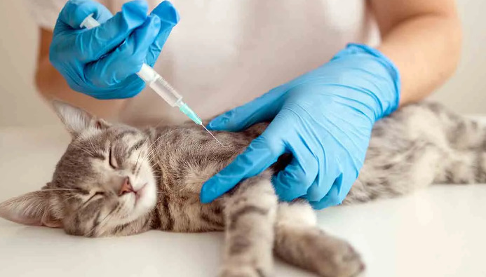
Fluid Analysis: Helps distinguish FIP effusion from other causes
Thus, diagnosis by exclusion integrates multiple lines of evidence—clinical suspicion, laboratory data, imaging findings, and response (or lack thereof) to therapy—while alternative diagnoses are actively ruled out. In high-suspicion scenarios, an exclusionary approach offers a pragmatic path, but challenges remain in ambiguous cases.
Modern Diagnostic Approaches: Beyond Exclusion
Recent advances are shifting the paradigm, supplementing — but not replacing — exclusionary diagnosis. Several tools are transforming the landscape:
Molecular Testing: PCR
Polymerase chain reaction (PCR) technology allows detection of FCoV RNA and, sometimes, FIP-associated mutations. PCR can be performed on blood, effusions, or tissue samples, raising hopes for definitive diagnosis. However, positive results confirm coronavirus presence, not necessarily FIP pathology. Mutation-specific PCRs (such as those targeting the M1058L mutation) can increase specificity but are not absolute.
Immunohistochemistry (IHC)
This approach stains tissue or fluid samples for viral antigen within macrophages. A positive result is regarded as strong evidence for FIP when contextualized with clinical findings. IHC, however, requires tissue, usually obtained via biopsy or at necropsy—a practical limitation.
Effusion Analysis: Rivalta Test and Biochemistry
In cases of wet FIP, the Rivalta test offers an inexpensive, bedside method to distinguish FIP exudate from other effusions. Positive tests point toward FIP, especially alongside classical clinical and laboratory hallmarks.
Combining Results for a Presumptive Diagnosis
Present-day FIP diagnosis hinges on the "weight of evidence" approach. No single gold standard exists that perfectly discriminates FIP from mimics. Veterinarians weigh history, risk factors (age, environment), clinical signs, lab findings, imaging features, and molecular results, seeking convergence upon a plausible diagnostic conclusion.
For many practitioners, exclusion remains essential—especially in settings where advanced diagnostics are inaccessible or equivocal. The rise of molecular diagnostics adds confidence but hasn’t removed the need for clinical judgment and systematic elimination of similar pathologies.
Distinguishing FIP from Related Conditions
FIP vs. Other Effusive Diseases
Abdominal or pleural fluid in cats may result from heart disease, bacterial infections, neoplasia, trauma, or FIP. Fluid analysis (cytology, protein concentration, Rivalta test), imaging, and serology are used to rule out alternative causes. In FIP, fluids are typically straw-colored, protein-rich, and cellular.
FIP vs. Lymphoma
Lymphoma, particularly abdominal and multicentric variants, can closely mimic FIP symptoms and imaging findings. Cytology from masses or fluids and advanced imaging (CT, ultrasound) often distinguish these, but tissue biopsy may be necessary.
FIP vs. Bacterial Peritonitis
Bacterial infections usually produce septic effusions with degenerative neutrophils and bacteria. FIP effusions are typically sterile. Culture and cytology differentiate these entities.
FIP vs. Other Infectious Diseases (e.g., Toxoplasmosis, Mycobacteriosis)
These can generate granulomatous lesions and fever. PCR and serology, alongside histopathological analysis, help clarify these diagnoses.
Limitations of Diagnosis by Exclusion
While exclusion remains central, pitfalls exist. Over-reliance on this approach may lead to misdiagnosis, premature euthanasia, or missed therapeutic opportunities. Molecular and immunohistochemical techniques, when available, can mitigate uncertainty, but have their own shortcomings—false negatives (especially if sample quality is poor or viral load low), and nonspecific positives (especially in endemic areas).
The Role of Clinical Suspicion and Pattern Recognition
Despite technological strides, the experience and intuition of the clinician remain at the heart of FIP diagnosis. Certain "red flag" features reliably prompt suspicion:
Persistent, fluctuating fever unresponsive to antibiotics
Young age (especially under two years old)
Multi-cat environment history
Effusions with high protein, low cellularity
Typical CBC/biochemical shifts (hyperglobulinemia, etc.)
These findings, integrated with diagnostic results and a process of elimination, help guide clinical action.
Diagnostic Algorithms: Integrating Old and New
To streamline FIP diagnosis, many veterinary centers and academic experts recommend diagnostic algorithms. These combine:
Initial screening: History, physical exam, CBC, biochemistry
Imaging: Detecting effusions, organ changes
Fluid/tissue analysis: Rivalta test, cytology, protein content
Molecular/IHC assays: PCR and latency tests as indicated
Each step refines the diagnostic probability, progressively ruling in or out alternative causes, isolating the likelihood of FIP.
Emerging Diagnostic and Treatment Horizons
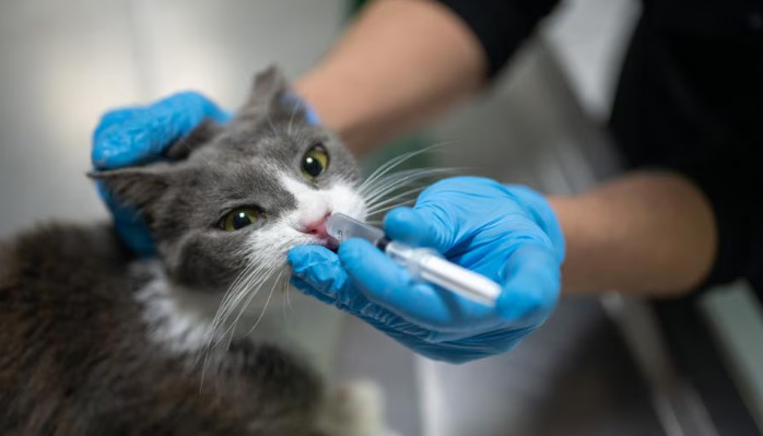
Recent years have witnessed dramatic advances in FIP therapy—most notably, antiviral medications like GS-441524 and its commercial derivatives, which can induce remission in many cases previously considered hopeless. Alongside this, diagnostic tools are improving, with novel PCR mutations, point-of-care assays, and tissue-specific markers under study.
As effective treatments emerge, the imperative for rapid, reliable diagnosis intensifies. More accurate discriminators are necessary, not only to confirm FIP but to avoid unnecessary application of costly treatments to non-FIP cases.
Summary Table: FIP Diagnostic Tools and Their Strengths/Limitations
| Diagnostic Method | Strengths | Limitations |
||-|-|
| Serology (Ab/Ag) | Rapid, accessible | Poor specificity; high coronavirus prevalence |
| Fluid analysis (Rivalta, etc)| Quick, useful for effusions | May misclassify; overlaps with other diseases |
| PCR (generic coronavirus) | Direct viral detection | Can't distinguish FIP vs. benign infection |
| Mutation-specific PCR | Increased specificity | May miss unknown mutations; not universally available|
| Immunohistochemistry (IHC) | High specificity | Needs tissue; invasive, sometimes not feasible|
| Imaging (US/X-ray/CT) | Details on effusions/masses | Non-specific; needs skilled interpretation |
| Clinical suspicion/exclusion | Integrates all available info | Subjective; risk of diagnostic error |
The Takeaway on FIP Diagnosis by Exclusion
FIP’s diagnosis remains a nuanced craft that blends exclusionary reasoning with sophisticated laboratory tools—when such resources are available. In the absence of a definitive, universally accessible diagnostic test, exclusion provides a logical framework to guide clinical decision-making, protect against misdiagnosis, and ensure that life-saving treatments reach the right patients. As science advances, new tests may one day supplant exclusion as the gold standard. Until then, the best path is careful, methodical reasoning, informed by the latest evidence and bolstered by innovation.
References
1. Pedersen NC. A review of feline infectious peritonitis virus infection: 1963–2008. J Feline Med Surg.
2. Tasker S, Dyer R. Feline infectious peritonitis: the diagnosis dilemma. Vet J.
3. Kipar A, Meli ML. Feline infectious peritonitis: Still an enigma? Vet Pathol.
4. Addie DD, Toth S, et al. Feline coronavirus, sequences in FIP diagnosis. J Feline Med Surg.
5. Hartmann K. Feline Infectious Peritonitis: advances in diagnosis and therapy. Vet Clin North Am Small Anim Pract.
6. Felten S, Weigner K. Diagnostics and laboratory tests for FIP. Vet Clin North Am Small Anim Pract.
7. Norris JM, et al. Clinical and laboratory findings associated with FIP effusions. Aust Vet J.
8. Stranieri A, et al. Markers and mutations for FIP diagnosis: recent evidence. J Vet Diagn Invest.
9. de Groot-Mijnes JD, et al. Threats and new directions for FIP testing. Vet Microbiol.
10. Chang HW, et al. Recent advances in molecularly targeted diagnosis for feline infectious peritonitis. Veterinary Research.