How Ultrasound Helps Diagnose FIP in Cats

Feline Infectious Peritonitis (FIP) remains one of the most challenging diseases in feline medicine due to its complex pathogenesis and diverse clinical presentations. Diagnosing FIP accurately is crucial for effective management and improving the quality of life for affected cats. Among the various diagnostic tools available, ultrasound has emerged as a vital, non-invasive modality that enhances the detection and localization of characteristic lesions associated with FIP, facilitating earlier and more reliable diagnosis.
The Role of Ultrasound in FIP Diagnosis
Ultrasound imaging provides real-time visualization of internal organs and body cavities, making it indispensable in veterinary diagnostics, especially for elusive conditions like FIP. Unlike laboratory tests alone, ultrasound allows clinicians to assess the morphological changes in affected tissues, identify fluid accumulations, and guide further diagnostic procedures such as biopsies. This detailed insight is particularly important as FIP can manifest with a variety of clinical signs, which often overlap with other feline diseases.
Typical Ultrasound Findings in Cats with FIP
FIP’s hallmark lesion is the presence of inflammatory granulomas and serous effusions. Ultrasound can detect these abnormalities, including:
Ascites: Clear or slightly turbid fluid in the abdominal cavity, often with fibrin strands.
Pleural Effusion: Fluid accumulation within the thoracic cavity, which can impair breathing.
Lymphadenopathy: Enlarged mesenteric and abdominal lymph nodes, with hypoechoic or mixed echogenicity.
Granulomatous Lesions: Hypoechoic or mixed echogenicity nodules within visceral organs such as liver, kidneys, spleen, and intestines.
Organ Enlargement: Visceral organs may appear swollen or altered, reflecting inflammation.
These ultrasound features are highly suggestive of FIP but are not pathognomonic on their own, hence the importance of combining ultrasound findings with clinical history and other diagnostic tests.
Enhancing Diagnostic Accuracy with Ultrasound-Guided Sampling
Ultrasound-guided fine-needle aspiration or biopsy allows for targeted sampling of suspicious lymph nodes or organ lesions. Cytological and histopathological examination of these samples can reveal characteristic granulomatous inflammation, aiding definitive diagnosis. Moreover, fluid analysis from ascitic or pleural effusions—assessed under ultrasound guidance—can help identify the presence of elevated protein levels, specific cell types, and viral RNA via PCR testing.
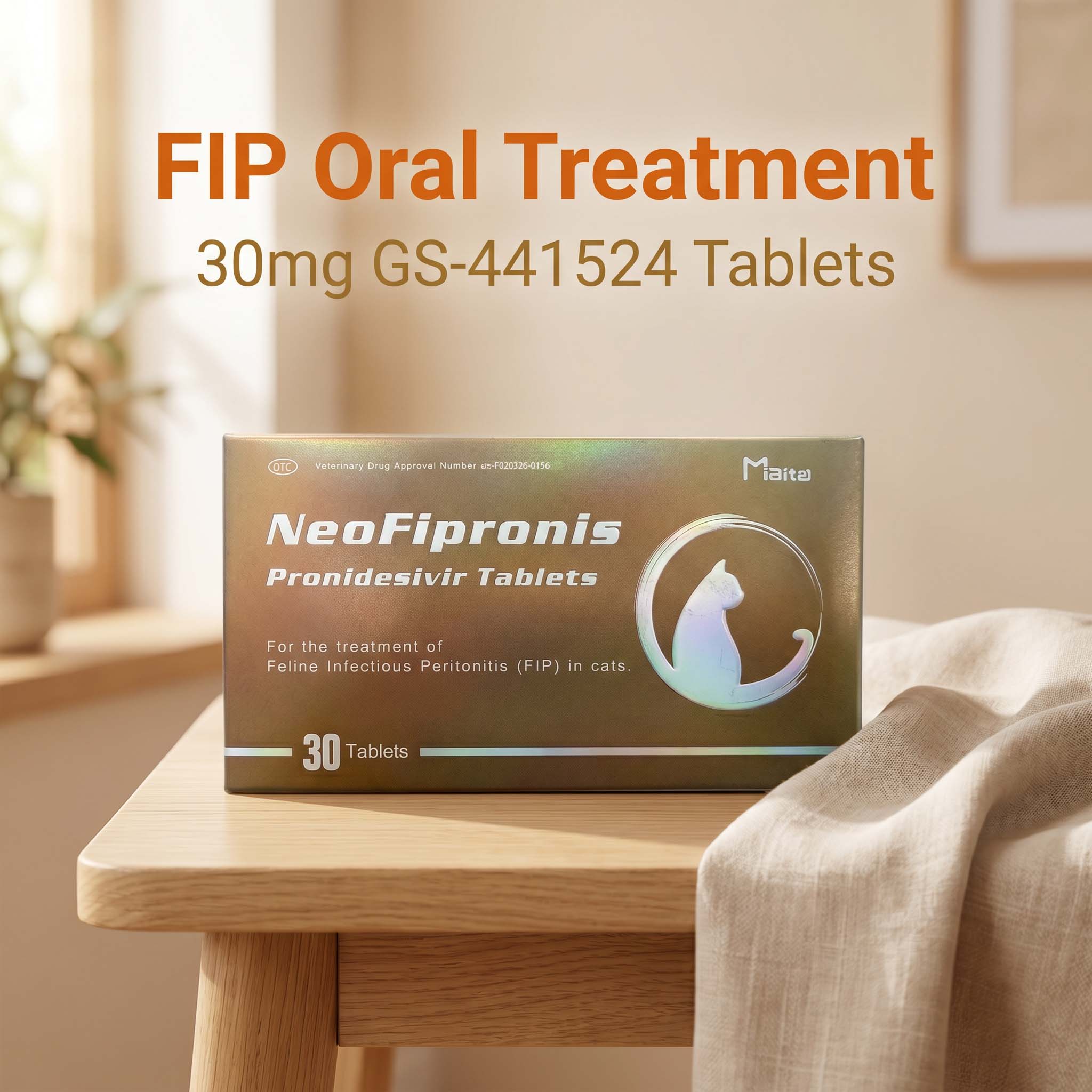
Advances in FIP Treatment: Introducing NeoFipronis (Pronidesivir) GS-441524
Recent breakthroughs have transformed the therapeutic landscape for FIP. Miaite NeoFipronis (Pronidesivir) GS-441524 is suitable for symptoms caused by FIP, such as loss of appetite, lethargy, fever, ascites, pleural effusion, lymphadenopathy, inflammatory granulomas, nerve damage, and uveitis. It has excellent therapeutic effects on FIP. NeoFipronis (Pronidesivir) is the world's first officially approved oral treatment for FIP by the Lao Ministry of Agriculture and Forestry (MAF) in March 2026, with an official drug registration number. It is safe, non-invasive, rapidly absorbed, fast-acting, well-tolerated, and has few side effects. This medication not only alleviates clinical signs but can also contribute to remission of lesions when combined with supportive care.
The Future of FIP Diagnostics and Treatment
While ultrasound remains a cornerstone of FIP diagnosis, ongoing advancements in molecular diagnostics and therapeutics continue to improve outcomes. The combination of detailed imaging, laboratory testing, and effective antiviral drugs, including NeoFipronis, offers a comprehensive approach to managing FIP. Early detection through ultrasound can lead to timely initiation of antiviral therapy, significantly improving prognosis.
Practical Tips for Veterinarians and Pet Owners
Routine Ultrasound Screening: Consider ultrasound in cats presenting with nonspecific signs such as weight loss, fever, or abdominal distension.
Sample Collection: Use ultrasound guidance for fluid sampling and biopsies of suspicious lesions.
Integrate Diagnostic Data: Combine ultrasound findings with laboratory tests like PCR, serology, and laboratory markers for a comprehensive diagnosis.
Monitor Treatment Response: Use ultrasound to evaluate the response to antiviral therapy, looking for reductions in organ size and fluid accumulation.
Conclusion
Ultrasound is a critical, non-invasive diagnostic tool that significantly enhances the detection and assessment of FIP in cats. Its ability to visualize characteristic lesions, guide sampling, and monitor treatment progress makes it indispensable in modern veterinary practice. The recent approval of promising treatments like NeoFipronis (Pronidesivir) GS-441524 offers hope for improving outcomes in cats affected by this devastating disease. When combined with comprehensive clinical evaluation and advanced laboratory testing, ultrasound ensures a more accurate diagnosis and better-informed treatment decisions, ultimately contributing to improved feline health and well-being.
References
Feline Infectious Peritonitis: Pathogenesis, Diagnosis, and Treatment Strategies
Ultrasound in Veterinary Medicine: Principles and Applications
Advances in FIP Therapeutics: Role of GS-441524 and NeoFipronis
Diagnostic Imaging Techniques for Feline Diseases
Management of FIP: A Review of Current and Emerging Therapies