How Is Feline Infectious Peritonitis (FIP) Usually Diagnosed

Feline Infectious Peritonitis (FIP) is a complex and often fatal disease caused by certain strains of the feline coronavirus (FCoV). While many cats are infected with FCoV, only a small percentage develop FIP, making diagnosis challenging. Veterinary professionals rely on a combination of clinical signs, laboratory tests, imaging techniques, and, occasionally, post-mortem examinations to confirm the presence of FIP.
Understanding the Disease and Its Challenges in Diagnosis
FIP manifests in two main forms: the effusive (“wet”) form and the non-effusive (“dry”) form, each presenting different clinical signs. The 'wet' form typically causes fluid accumulation within body cavities, such as the abdomen or chest, whereas the 'dry' form involves granulomatous lesions in organs like the liver, kidneys, or lymph nodes. The variable presentation complicates diagnosis because symptoms often overlap with other diseases like cancer, bacterial infections, or liver failure.
The crucial aspect of diagnosis lies in differentiating FIP from other conditions with similar clinical signs. Historically, definitive diagnosis has been difficult without invasive procedures or post-mortem analysis. Recent advances, however, have improved the ability to diagnose FIP ante-mortem with a higher degree of confidence.
Clinical Examination and History
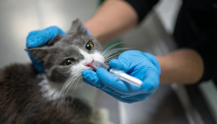
Initial assessment begins with a thorough clinical examination and detailed medical history. Veterinarians look for signs such as weight loss, lethargy, fever unresponsive to antibiotics, and characteristic fluid buildup. Medical history might include exposure to multi-cat environments or previous outbreaks of feline coronavirus infection. These details guide clinicians towards suspecting FIP, especially in young or immunocompromised cats.
Laboratory Tests
Laboratory analysis forms the cornerstone of FIP diagnosis. The following tests are commonly employed:
Complete Blood Count (CBC): Often reveals a non-specific inflammatory response, with some cases showing anemia, lymphopenia, or leukocytosis.
Serum Chemistry Panel: May demonstrate elevated liver enzymes, hyperglobulinemia, or decreased albumin, resulting in a low albumin-to-globulin ratio, which is suggestive but not definitive for FIP.
Effusion Analysis: In cats with the wet form, fluid analysis is crucial. The fluid is typically straw-colored or yellowish, with high protein content (>3.5 g/dL) and low cellularity. Cytology reveals a mild neutrophilic or mixed inflammatory infiltrate without bacteria, supporting an immune-mediated process.
Serological and Molecular Tests
While antibody titers against feline coronavirus are sensitive indicators of exposure, they are not specific for FIP since many healthy cats may carry FCoV without developing FIP. Conversely, high titers are suggestive but not conclusive, as some healthy cats also show elevated levels.
Polymerase Chain Reaction (PCR) testing detects viral RNA in blood, tissue, or effusions. Its sensitivity is high, but it can also detect non-pathogenic virus, complicating interpretation. To increase diagnostic accuracy, detecting viral RNA within macrophages of effusions or tissues via PCR is considered more indicative of FIP.
Imaging Techniques
Radiographs and ultrasound are valuable adjuncts. Ultrasound can identify characteristic changes such as thickened organ walls, granulomatous lesions, or abdominal effusion. On thoracic radiographs, pleural effusions or granulomas may be observed. While these findings support FIP suspicion, they are not definitive on their own.
Histopathology and Post-mortem Diagnosis
When feasible, tissue biopsy or necropsy remains the gold standard for confirming FIP. Histopathological examination reveals granulomatous inflammation of affected organs with FIP-specific lesions. Immunohistochemistry (IHC) staining for feline coronavirus within macrophages confirms the diagnosis. However, this approach is invasive and often only performed in post-mortem analyses, limiting its use in live patients.
Emerging Diagnostic Tests and Future Directions
Recent developments include specific in vitro assays such as the FIP virus-specific antibody test and novel molecular techniques that improve diagnostic confidence. Despite advancements, no single test definitively diagnoses FIP in all cases, and a combination of clinical, laboratory, and imaging findings remains essential.
Integrating the Diagnostic Process
Effective diagnosis of FIP involves integrating multiple lines of evidence:
1. Clinical signs consistent with FIP
2. History of exposure to feline coronavirus
3. Fluid analysis supportive of FIP
4. Laboratory findings indicating systemic inflammation
5. Imaging studies revealing characteristic lesions
6. Molecular detection of viral RNA in tissues or fluids
The diagnostic process requires careful interpretation of all data points, acknowledging limitations inherent to each method. In complex cases, consultation with veterinary specialists, especially in infectious disease or pathology, can aid in diagnosis.
Conclusion
Diagnosing FIP is a nuanced process demanding a multi-modal approach. No single test guarantees accuracy, but combining clinical assessment with laboratory, imaging, and increasingly, molecular diagnostics can lead to a probable or confirmed diagnosis. Ongoing research promises to improve diagnostic accuracy further, aiding in earlier detection and better management of this challenging feline disease.
References
1. Pedersen, N. C. (2014). Feline Infectious Peritonitis: Dogma Disproved. Journal of Feline Medicine and Surgery, 16(3), 201–209.
2. Addie, D. D., & Jarrett, O. (2004). Feline Coronavirus Infection. Feline Medicine and Surgery, 2nd Edition. Saunders Ltd.
3. Hartmann, K. (2011). Feline Infectious Peritonitis. The Veterinary Clinics of North America: Small Animal Practice, 41(2), 319–328.
4. Pesteanu-Somogyi, L., & Hegedüs, J. (2015). Diagnosis and Management of Feline Infectious Peritonitis. Veterinary Medicine International, 2015, 1–9.
5. Kipar, A., et al. (2006). Pathogenesis of Feline Infectious Peritonitis. Journal of Comparative Pathology, 135(2–3), 125–144.