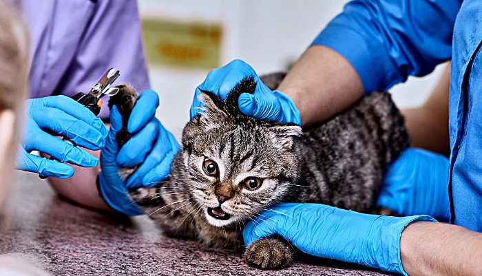
Is FIP Only Detectable Through Careful Observation

Feline Infectious Peritonitis (FIP) is one of the most challenging diseases faced by cat owners and veterinarians. Caused by a mutated strain of feline coronavirus (FCoV), FIP remains enigmatic due to its diverse clinical presentation and the lack of a single, definitive diagnostic test. The complexities of FIP have led many to wonder whether careful observation alone can reveal its presence, or if more advanced diagnostic procedures are essential. This article explores the clinical, laboratory, and imaging aspects of FIP detection, clarifies the limitations of observation, and reviews the latest diagnostic approaches.
Understanding FIP: What Makes It Difficult to Diagnose?
The pathogenesis of FIP begins when a relatively benign coronavirus infects a cat, usually without causing severe illness. Most cats will experience mild diarrhea or respiratory symptoms, or remain asymptomatic. However, in some cases, the virus mutates within the cat and gains the ability to infect macrophages, triggering an aberrant immune response that results in FIP. This mutated form is not contagious; instead, the risk lies in exposure to the original, widespread coronavirus.
FIP presents in two general forms: the “wet” (effusive) and “dry” (non-effusive). The wet form leads to accumulation of fluid within the abdomen or chest, causing visible swelling, respiratory distress, and lethargy. The dry form is marked by granuloma formation and organ dysfunction without significant effusion, resulting in more subtle clinical signs such as neurological symptoms, ocular changes, unthrifty coat, and weight loss.
Careful Observation: What Can It Reveal?
Careful observation can alert owners and veterinarians to the possibility of FIP but is rarely sufficient for definitive diagnosis. Typical warning signs include:
Persistent fever unresponsive to antibiotics
Sudden or progressive weight loss
Lethargy and decreased appetite
Jaundice or pale gums
Distended abdomen (especially in the wet form)
Trouble breathing from fluid in the chest
Neurological signs: changes in gait, head tilt, nystagmus, or seizures
Ocular signs: inflammation, changes in pupil size, retinal detachment
These signs are suggestive but far from specific; many other diseases, such as lymphomas, other infectious diseases (toxoplasmosis, cryptococcosis), nutritional disorders, and metabolic illnesses can mimic FIP’s presentation. This overlap makes careful observation essential for raising clinical suspicion, but not for confirming or excluding FIP.
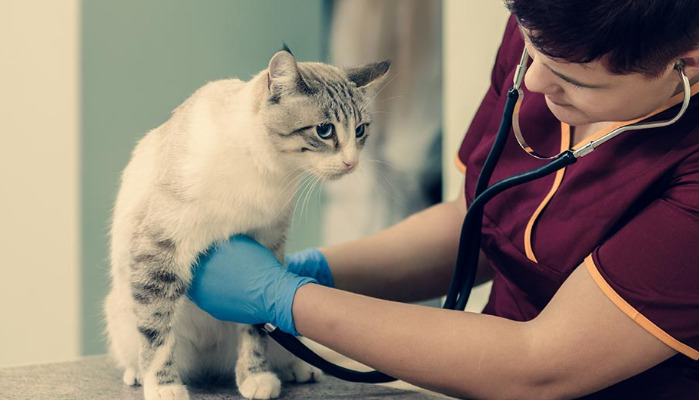
Diagnostic Laboratory Investigations
Once FIP is suspected based on clinical observation, laboratory testing plays a crucial role in diagnosis. Routine tests may include:
Complete Blood Count (CBC):
Lymphopenia (low lymphocyte count)
Neutrophilia (high neutrophil count)
Non-regenerative anemia
Serum Biochemistry:
Hyperglobulinemia (elevated globulins)
Low albumin-to-globulin ratio (<0.8 often seen in FIP)
Elevated liver enzymes or bilirubin
Elevated total protein
None of these findings is exclusive to FIP, but the constellation of abnormalities, especially in conjunction with compatible clinical signs, raises suspicion.
Effusion Analysis:
In effusive FIP, the fluid removed from the abdomen or chest is typically straw-colored, viscous, and protein-rich. Its analysis often shows high protein content (>3.5 g/dl), low cellularity, and a predominance of neutrophils and macrophages.
Rivalta Test:
This simple, in-clinic test exposes effusion fluid to acetic acid; a positive result supports the possibility of FIP, though not definitively, and false positives/negatives can occur.
Imaging: X-rays and Ultrasound
Imaging studies complement physical observation and laboratory testing. Abdominal and thoracic radiographs can reveal fluid accumulation, organ enlargement, and lymphadenopathy. Ultrasound is especially useful to localize fluid, assess organ structure, and guide sample collection for further analysis.
The “dry” form of FIP can cause granulomas in organs such as kidneys, liver, lymph nodes, and even the central nervous system. While imaging can detect these changes, they remain non-specific and do not singularly confirm FIP.
Advanced Diagnostics: Molecular and Immunological Testing
Definitive confirmation of FIP often requires detection of the mutated virus or its genetic material in affected tissues, fluids, or blood.
RT-PCR (Reverse Transcription Polymerase Chain Reaction):
PCR can detect FCoV RNA in effusion fluids, tissues, or blood but does not always distinguish between the benign enteric form and the mutated FIP-causing variant. Advanced PCR tests seek mutations associated with virulence, but their sensitivity and specificity can vary.
Immunohistochemistry (IHC):
Staining tissue biopsies for FCoV antigens inside macrophages is considered the gold standard for FIP but requires invasive sample collection and specialized labs.
Serology:
Antibody tests identify exposure to enteric FCoV but cannot discern whether a cat has developed FIP. Most cats exposed to FCoV do not get FIP.
Gene Sequencing:
Some reference labs can sequence viral RNA from fluid or tissue samples to identify mutations strongly associated with FIP. However, this is expensive and not universally available.
Role of Clinical Judgment
The complexity and variability of FIP means that, strictly speaking, observation alone cannot “detect” FIP in most cases. It serves as the critical first step — prompting further evaluation when signs and symptoms are concerning. Ultimately, a diagnosis is a synthesis of the cat’s history, physical examination, laboratory and imaging findings, and results of advanced testing where available.
A veterinarian’s clinical judgment, especially when familiar with FIP’s protean manifestations, remains vital. They integrate findings to arrive at the most probable diagnosis and exclude alternative causes. In some cases, unfortunately, the diagnosis is only confirmed post-mortem through tissue examination.
Current and Emerging Techniques in FIP Detection
New diagnostic approaches are being evaluated as understanding of FIP grows. Commercial FCoV mutation PCRs, improved imaging modalities, and next-generation sequencing may one day improve the speed and accuracy of FIP detection.
Liquid biopsies (testing for circulating FCoV mutation signatures in blood) and improved point-of-care molecular tests aim to support vets and owners in timely decision making for both diagnostic and therapeutic purposes.
However, cost, availability, and technical requirements limit widespread adoption of these tools. Thus, the combination of careful observation with classical and advanced diagnostics remains the cornerstone of FIP detection.
Why FIP Is Not Only Detectable by Observation
Cats with subtle or atypical signs — neurological changes, behavioral abnormalities, or passing episodes of lethargy — may escape observation-based diagnosis. Many systemic disorders can present with lethargy, fever, or ascites without being FIP.
For example, heart disease may cause fluid accumulation, and certain cancers can mimic both wet and dry FIP. Kidney or liver failure, autoimmune disease, and other infections enter the differential diagnosis. Without laboratory confirmation, practitioners risk misdiagnosis and inappropriate or delayed therapy.
Moreover, the emotional and ethical weight of a potential FIP diagnosis pushes clinicians toward a comprehensive approach. As devastating as FIP is, a diagnosis based solely on observation could miss treatable causes, or mislead owners into thinking the situation is hopeless.
Importance of Timely Diagnosis
Recent breakthroughs in therapeutic options for FIP (such as antiviral drugs GS-441524 and remdesivir, which may become more widely available in the future in the United States depending on ongoing research and regulation) mean that early and accurate identification of FIP is more crucial than ever. Observational findings must trigger timely diagnostic testing, because cats who receive treatment in the earlier stages of disease respond best.
Not only does early diagnosis enable better survival rates with these emerging drugs, but it also guides isolation protocols, informs owner decision making, and prevents unnecessary euthanasia.
Conclusion: Integration Is Essential
The key to FIP detection lies not in one method, but in integration — combining careful clinical observation with thorough laboratory, imaging, and molecular diagnostics. Observation is a vital first step, alerting caregivers and clinicians to danger, but it cannot stand alone.
All in all, while observation is indispensable in identifying cats at risk or raising clinical suspicion of FIP, it does not suffice for reliable detection. Cats with suspicious signs require a targeted diagnostic approach, and emerging molecular tests may soon enhance the accuracy and speed of FIP diagnosis, improving outcomes for this devastating feline disease.
References
1. Pedersen, N.C. (2009). A review of feline infectious peritonitis virus infection: 1963–2008. Journal of Feline Medicine and Surgery, 11(4), 225-258.
2. Addie, D.D., et al. (2022). Feline infectious peritonitis: ABCD guidelines on prevention and management. Journal of Feline Medicine and Surgery, 24(4), 313–334.
3. Kipar, A., & Meli, M.L. (2014). Feline infectious peritonitis: Still an enigma? Veterinary Pathology, 51(2), 505–526.
4. Hartmann, K. (2005). Feline infectious peritonitis. Veterinary Clinics of North America: Small Animal Practice, 35(1), 39-79.
5. Felten, S., & Hartmann, K. (2019). Diagnosis of feline infectious peritonitis: A review of the current literature. Viruses, 11(11), 1068.
6. Dempsey, S.M., & Ewing, P.J. (2011). Feline infectious peritonitis: Diagnostic dilemmas. Compendium: Continuing Education for Veterinarians, 33(8), E1-E8.
7. Chang, H.W., et al. (2012). Detection of feline coronavirus with mutations in the spike gene associated with feline infectious peritonitis in cat tissues and body fluids. Journal of Clinical Microbiology, 50(8), 2648-2652.
8. Fischinger, K., & Korn, K. (2023). Diagnostics for feline infectious peritonitis: recent advances and challenges in field conditions. Pathogens, 12(1), 56.
9. Drut, A., et al. (2021). Feline infectious peritonitis: diagnostic protocol update and review. Veterinary Sciences, 8(9), 190.
10. Vennema, H. (1999). Feline infectious peritonitis: an update on pathogenesis and diagnostics. Advances in Experimental Medicine and Biology, 473, 345–360.