How Is Dry FIP Diagnosed in Cats

Feline Infectious Peritonitis (FIP) remains one of the most challenging diagnoses in feline medicine. FIP, caused by a mutant strain of feline coronavirus (FCoV), presents in two major forms: wet (effusive) and dry (non-effusive). While wet FIP often exhibits more obvious clinical signs due to fluid accumulation, dry FIP proves trickier to identify because its manifestations are subtle and diverse. This article aims to unravel the diagnostic complexities of dry FIP, offering a detailed roadmap for veterinarians and cat owners based in the US.
What is Dry FIP?
Unlike wet FIP, which is marked by fluid buildup in the abdomen or chest, dry FIP primarily affects organs through granulomatous lesions—localized clusters of inflammation. These lesions may appear in the kidneys, liver, lymph nodes, eyes, and even the brain, leading to a wide variety of symptoms depending on the organs involved. The biphasic nature and variability in dry FIP's clinical picture complicate its recognition, especially in initial stages.
Clinical Presentation: The Crucial Clues
Recognizing dry FIP starts with a thorough assessment of the cat’s symptoms. While there is no single sign diagnostic for the disease, several non-specific indicators should raise suspicion:
Persistent fever unresponsive to antibiotics
Lethargy and weight loss
Anorexia
Eye changes (anterior uveitis, retinal detachment, keratic precipitates)
Neurological signs (ataxia, seizures, behavioral changes)
Enlarged lymph nodes or abnormal kidney/liver palpation
Eye and neurological symptoms are more strongly correlated with dry FIP than the wet form. Often, a careful medical history will reveal that the cat is young (less than two years old), comes from a multi-cat household or shelter, and has experienced chronic stress.
Stepwise Diagnostic Approach
1. Medical History and Physical Examination
The foundation of dry FIP diagnosis lies in combining clinical intuition with careful observation. Age, housing environment, previous health issues, and exposure history are critical. The veterinarian conducts a hands-on exam, looking for palpable kidney enlargement, irregular liver margins, and inspecting the eyes for inflammation.
2. Laboratory Testing
Dry FIP cannot be definitively confirmed by routine laboratory tests, but several findings provide strong supporting evidence. Key laboratory clues include:
Complete Blood Count (CBC): Mild to moderate non-regenerative anemia; lymphopenia (low lymphocyte count), and neutrophilia (high neutrophil count).
Serum Biochemistry: Hyperglobulinemia (especially elevated γ-globulins), decreased albumin/globulin ratio (below 0.8), mild increases in liver enzymes, and possible azotemia.
FCoV Antibody Titers: High titers may suggest exposure, but they do not distinguish between FIP and harmless FCoV infection. Titers alone can't confirm FIP.
Acute Phase Proteins: High α-1 acid glycoprotein levels support the suspicion of FIP.
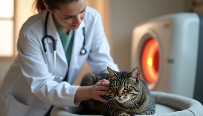
3. Imaging Studies
Ultrasound and Radiography: Imaging can reveal organomegaly, abdominal masses, lymphadenopathy, or abnormal tissue consistency. Although neither is pathognomonic, they help guide needle aspirates or biopsies.
Ocular Examination: Slit-lamp and fundoscopic analysis often reveals anterior uveitis or chorioretinitis. These signs, when paired with systemic symptoms, are suggestive of dry FIP.
4. Cytology and Histopathology
Fine Needle Aspirate: Samples from lymph nodes, kidneys, and affected organs may reveal pyogranulomatous inflammation.
Tissue Biopsy: The gold standard for FIP diagnosis. Histopathological evaluation demonstrates granulomatous (macrophage-dominated) inflammation, vasculitis, and necrosis. Immunohistochemical staining for FCoV antigen within macrophages is highly specific for FIP.
Molecular Diagnostics: Refining Accuracy
1. Reverse Transcriptase Polymerase Chain Reaction (RT-PCR):
RT-PCR detects viral RNA from tissue, effusions, or cerebrospinal fluid. In dry FIP, samples may be taken from affected organs or lymph nodes. However, RT-PCR has limitations—it can be negative despite disease presence due to low viral loads, or positive in healthy FCoV carriers.
2. Immunohistochemistry:
Testing for viral antigen within granulomas confirms that the inflammation is caused by FCoV, not other agents. This test is considered definitive if positive but requires tissue samples.
3. Advanced PCR Mutational Analysis:
Newer PCR assays investigate specific FCoV mutations (such as the M1058L mutation in the spike gene) that are more commonly associated with the FIP-causing strain. While not yet universally available, these assays are improving specificity.
Differentiating Dry FIP from Other Diseases
The absence of effusion means dry FIP can mimic dozens of other illnesses. Diseases often confused with dry FIP include:
Lymphoma or other tumors
Chronic bacterial or fungal infections
Toxoplasmosis
Immune-mediated diseases (like lupus or polyarthritis)
Amyloidosis
A careful stepwise approach, combining history, physical examination, laboratory results, and, when feasible, tissue diagnosis, remains the most effective way to rule in or rule out dry FIP.
Key Diagnostic Tests and Their Significance
1. Albumin:Globulin Ratio
An A:G ratio less than 0.8 increases suspicion for FIP. Values below 0.4 strongly suggest FIP in the right clinical context.
2. α-1 Acid Glycoprotein Levels
Elevated >1.5 mg/ml are highly suggestive of FIP but not exclusive.
3. Immunohistochemistry for FCoV Antigen
The only test that definitively confirms FIP upon demonstration of viral antigen within macrophages in pyogranulomatous tissue.
4. PCR Mutation Analysis
Confirms FIP-associated FCoV if positive from affected tissues.
Challenges and Limitations
Despite advances, many veterinary clinics lack access to sophisticated testing, especially immunohistochemistry and mutational PCR. Most diagnoses rely on clinical suspicion and exclusion of other diseases. The ethical dilemma of invasive diagnostics arises—many cats are too ill for surgical biopsies. Recent innovations in minimally invasive laparoscopic biopsies and improved imaging are assisting, but obstacles remain.
Special Considerations: Neurological and Ocular FIP
Neurological and ocular forms pose extra hurdles. When the nervous system is affected, symptoms can include seizures, ataxia, or behavioral changes. Ocular signs often present early; therefore, ophthalmological examination is pivotal. Diagnosis often depends on cerebrospinal fluid analysis, with increased protein and cell counts, along with RT-PCR or antibody testing of the fluid.
Emerging Diagnostic Techniques
Continued research aims to improve rapid, non-invasive diagnostics. Potential blood biomarkers, point-of-care PCR kits, and advanced imaging modalities (such as MRI for neurological involvement) offer hope for earlier and more accurate diagnoses. Whole-genome sequencing and proteomics may soon become part of routine screening in specialized centers.
Implications for Treatment and Prognosis
Accurate diagnosis is key—many promising therapies for FIP, including antiviral drugs like GS-441524 and remdesivir, are now available under compassionate protocols in the US. These treatments, however, are costly and require clear diagnosis to justify use. Misdiagnosis can result in inappropriate therapy and unnecessary euthanasia.
Preventive Strategies and Owner Guidance
While diagnosis is central, prevention remains crucial. Most cats exposed to FCoV will not develop FIP, but stress minimization, population control in shelters, rigorous hygiene, and early elimination of high-risk carriers have been shown to reduce incidence. Owners should report any persistent, unexplained symptoms to their veterinarian and pursue aggressive diagnostic testing if dry FIP is suspected.
The Role of the Veterinarian in Diagnosis
Veterinarians must balance the need for diagnostic certainty with the welfare and comfort of their feline patients. Empathy, communication, and transparency are essential when discussing possibilities and outcomes with owners. Diagnostic algorithms and continuing education ensure the latest developments are incorporated into practice.
Final Thoughts on Diagnostic Strategies
Diagnosing dry FIP is a complex interplay of art and science, demanding awareness of subtle symptoms, skilled interpretation of tests, and judicious use of invasive techniques. Emerging molecular and imaging technologies hold promise for future improvement, but currently, a multidisciplinary and cautious approach provides the best chance of timely and accurate diagnosis.
References
Sparkes, A. H., Gruffydd-Jones, T. J., Harbour, D. A., & Werrett, K. (1991). Feline infectious peritonitis: A review of clinical and diagnostic features. Journal of Feline Medicine and Surgery
Pedersen, N. C. (2014). Feline infectious peritonitis: an update. The Veterinary Journal
Tasker, S., & Malik, R. (2017). Feline infectious peritonitis: diagnosis and treatment update. Topics in Companion Animal Medicine
Kipar, A., & Meli, M. L. (2014). Feline infectious peritonitis: present status and future prospects. Veterinary Pathology
Felten, S., & Hartmann, K. (2019). Diagnosis of feline infectious peritonitis: a review of the current literature. Viruses
Dempsey, S. M., & Ewing, P. J. (2011). Feline infectious peritonitis: diagnostic challenges and current therapies. The Compendium: Continuing Education for Veterinarians
Norris, J. M., et al. (2005). Clinical and pathological findings associated with feline infectious peritonitis in Sydney, Australia: 42 cases (1990–2002). Australian Veterinary Journal