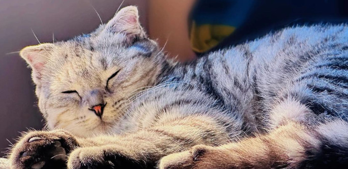
Eye Symptoms of Dry FIP in Cats

Feline Infectious Peritonitis (FIP) is an enigmatic and fatal disease affecting domestic cats worldwide. Caused by a mutation of feline coronavirus (FCoV), FIP typically presents two forms: effusive (wet) and non-effusive (dry). The dry form often involves neurological and ophthalmic signs, which can be challenging to recognize due to their variability and subtlety. For veterinarians, pet owners, and researchers, understanding ocular manifestations is crucial for timely diagnosis and intervention.
Pathophysiology of Dry FIP and Ocular Involvement
The dry form of FIP is characterized by granulomatous inflammation rather than the fluid accumulation seen in wet FIP. This immune-mediated response allows mutant FCoV to disseminate through macrophages, infiltrating organs such as the eyes, nervous system, and lymph nodes. In the eye, the uveal tract—including the iris, ciliary body, and choroid—is often targeted, resulting in a spectrum of ocular symptoms.
This infiltration leads to breakdown of the blood-ocular barrier, triggering inflammatory responses that can vary from mild to severe. The resulting tissue damage is responsible for most of the eye symptoms seen in dry FIP.
Common Ocular Symptoms in Dry FIP
Anterior Uveitis
Anterior uveitis, inflammation of the iris and ciliary body, is one of the most frequent ocular complications. Signs might include:
Redness (conjunctival hyperemia)
Cloudiness of the cornea (due to keratic precipitates)
Photophobia (light sensitivity)
Ocular pain (cats may squint or paw at the eye)
Change in iris color (iridial depigmentation)
These signs are not exclusive to FIP but their occurrence in young cats, especially with systemic illness, should raise concern.
Aqueous Flare
Aqueous flare refers to protein and inflammatory cell leakage into the anterior chamber, making it appear cloudy on slit-lamp examination. This symptom arises from increased permeability of inflamed vessels in the uveal tract.
Miosis and Irregular Pupil Shape
Inflammation can result in miosis (small pupil) or abnormally shaped pupils due to adhesions between the iris and lens (posterior synechiae). These signs are subtle but indicative of chronic uveitis commonly seen in FIP.
Hyphema and Hypopyon
Some cats develop hyphema (blood in the anterior chamber) or hypopyon (white blood cells/pus in the anterior chamber). These findings, though rare, are associated with more aggressive ocular involvement.
Posterior Segment Manifestations
Chorioretinitis
Inflammation may extend to the choroid and retina. Chorioretinitis presents with:
Blurred retinal borders
Retinal detachments
Hemorrhage or exudates in the posterior segment
Decreased vision or blindness
Fundoscopy can help detect subtle signs, although affected cats may not show obvious vision loss early on.
Retinal Vessel Changes
Feline FIP-related vasculitis often affects the retinal vessels, resulting in:
Perivascular cuffing (inflammatory cells surrounding blood vessels)
Hemorrhages
Vessel attenuation
These findings point to systemic vascular involvement.
Other Ocular Signs
Keratic Precipitates
Small, whitish deposits on the corneal endothelium resulting from inflammatory cells settling out of the aqueous humor are characteristic. These are visible during slit-lamp biomicroscopy.
Glaucoma
Chronic inflammation can block aqueous drainage pathways, leading to increased intraocular pressure (secondary glaucoma). Signs include:
Enlarged, cloudy eye
Pain and vision loss
Corneal edema
Glaucoma in FIP is often refractory and may progress rapidly.
Systemic and Neurological Associations
Cats with ocular FIP often have multisystemic disease. Neurological signs, including ataxia, seizures, or altered mentation, are frequently present alongside eye symptoms. Fever, weight loss, lethargy, and anorexia are common. These associations can aid the clinician in recognizing FIP as the underlying cause of the ocular pathology.
Diagnostic Challenges
Clinical Examination
Ophthalmic examination should be thorough, including slit-lamp and indirect ophthalmoscopy. While findings can be suggestive, ocular lesions alone are not pathognomonic for FIP.
Laboratory Testing
PCR and Serology: Testing ocular fluids (aqueous humor, vitreous) for FCoV RNA via PCR can help increase diagnostic certainty.
Cytology: Analysis of ocular fluids may reveal neutrophils or macrophages, but not always specific for FIP.
Blood Work: Hyperglobulinemia, lymphopenia, and elevated alpha-2 globulin fractions are commonly seen in cats with FIP.
Imaging: Advanced imaging (ocular ultrasound, CT) can help evaluate structural changes.
Histopathology
Definitive diagnosis requires biopsy or postmortem examination, revealing pyogranulomatous inflammation and viral antigen in ocular tissue.
Differential Diagnoses
Other diseases that mimic FIP’s ocular signs include:
Lymphoma (especially intraocular lymphoma)
Toxoplasmosis
Feline leukemia virus (FeLV) infections
Systemic fungal diseases (e.g., cryptococcosis)
Idiopathic uveitis
Careful consideration of age, history, clinical signs, and laboratory findings are critical in reaching an accurate diagnosis.
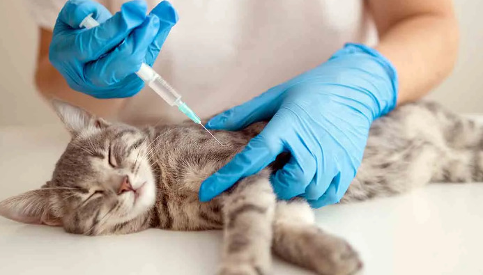
Treatment Strategies
Supportive Management
While FIP is historically considered fatal, supportive care—including nutritional support, pain management, and anti-inflammatories—can improve comfort and quality of life.
Targeted Therapies
Recent advances in antiviral drugs (e.g., GS-441524 analogs) have shown promise in extending survival and even resolving FIP. These therapies may improve ocular signs alongside systemic disease. Their access and legality may vary by region.
Ocular-Specific Treatments
Topical corticosteroids can help control uveitis, but systemic use must be weighed against possible immunosuppression. Glaucoma treatments (carbonic anhydrase inhibitors, beta-blockers) may be considered, though response is variable.
Prognosis
Historically, FIP had a grave prognosis, especially with ocular and neurological signs indicating disseminated disease. However, with new antiviral treatments, some cats show remarkable improvement. Early recognition and intervention remain key to increasing survival rates.
Conclusion
The ocular symptoms of dry FIP in cats represent a complex clinical challenge. Red eyes, vision loss, and changes in appearance should prompt a thorough diagnostic workup—especially in young, purebred cats. Advances in antiviral therapy offer new hope, but early intervention remains crucial. For veterinarians, being vigilant for ocular signs can make a significant impact on the outcome for cats affected by this devastating disease.
References
1. Hartmann, K. (2005). Feline infectious peritonitis. Veterinary Clinics of North America: Small Animal Practice, 35(1), 39-79.
2. Pedersen, N. C. (2009). A review of feline infectious peritonitis virus infection: 1963-2008. Journal of Feline Medicine and Surgery, 11(4), 225-258.
3. Andrew, S. E., et al. (1997). Ocular manifestations of systemic diseases in cats. Clinical Techniques in Small Animal Practice, 12(1), 45–54.
4. Montali, R. J., et al. (1995). Pathology of eye involvement in cats with FIP. Veterinary Pathology, 32(6), 501-506.
5. Paltrinieri, S., et al. (2020). Feline infectious peritonitis (FIP): New perspectives for diagnosis and management. Journal of Feline Medicine and Surgery, 22(10), 875-883.
6. Fischer, Y., & Ritz, S. (2014). Ocular disease associated with feline infectious peritonitis—a retrospective study of 55 cats. Journal of Feline Medicine and Surgery, 16(12), 992-998.
7. Felten, S., & Hartmann, K. (2019). Diagnosis of feline infectious peritonitis: A review of the current literature. Viruses, 11(2), 106.
8. Kipar, A., & Meli, M. L. (2014). Feline infectious peritonitis: Still an enigma? Veterinary Pathology, 51(2), 505-526.
9. Morrisey, J. (2021). Ocular manifestations of feline infectious peritonitis. Compendium: Continuing Education for Veterinarians, 43(4), E1-E6.
10. Dickinson, P. J., et al. (2020). Antiviral drug treatment of feline infectious peritonitis using GS-441524. Journal of Feline Medicine and Surgery, 22(4), 376-389.