Can Dry FIP Be Diagnosed Without Tissue Biopsy

Feline Infectious Peritonitis (FIP) poses profound challenges in feline medicine, particularly its elusive “dry” form. Caused by certain strains of feline coronavirus (FCoV), FIP can emerge either as effusive (wet) or non-effusive (dry). While wet FIP typically presents with characteristic fluid accumulation, making clinical identification more straightforward, dry FIP frequently lacks these hallmark features. This diagnostic conundrum has long made the disease one of the most feared and misunderstood pathologies in cats.
Historically, the gold standard for confirming dry FIP has been tissue biopsy and subsequent immunohistochemistry (IHC). Such invasive testing, however, is not always feasible due to the patient’s health, owner resources, and the practical difficulties in safely obtaining tissue samples. Accordingly, veterinarians and feline researchers continue to seek ways to reliably diagnose dry FIP without resorting to a tissue biopsy. This article explores current diagnostic strategies, their strengths and pitfalls, and what the evolving landscape reveals about the possibility of making this tough diagnosis without a scalpel.
What Is Dry FIP?
FIP is triggered when certain mutant forms of feline coronavirus (FCoV) gain the ability to replicate inside macrophages, leading to a progressive and nearly always fatal immune-mediated disease. In dry FIP, the infection prompts granulomatous inflammatory lesions in organs such as the kidneys, lymph nodes, liver, and sometimes the central nervous system or eyes. Unlike wet FIP, these cats rarely develop visible ascites or pleural effusion; thus, the diagnosis relies heavily on other clues, making it challenging without biopsy-confirmed evidence.
The Limitations of Biopsy
While tissue biopsy with IHC remains a definitive diagnostic tool, it can be risky or impossible for some cats. Cats with advanced dry FIP are often poor anesthesia candidates, and the location of lesions may make sampling impractical. Even fine-needle aspirates (FNA) may not yield sufficient tissue for firm conclusions.
Biopsy is expensive, time-consuming, and can cause further morbidity, particularly in weak cats. Some owners may decline for financial or ethical reasons. Thus, non-invasive diagnostic alternatives are increasingly essential in clinical practice.
Clinical Signs and Physical Examination
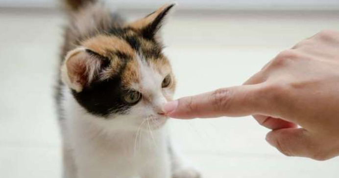
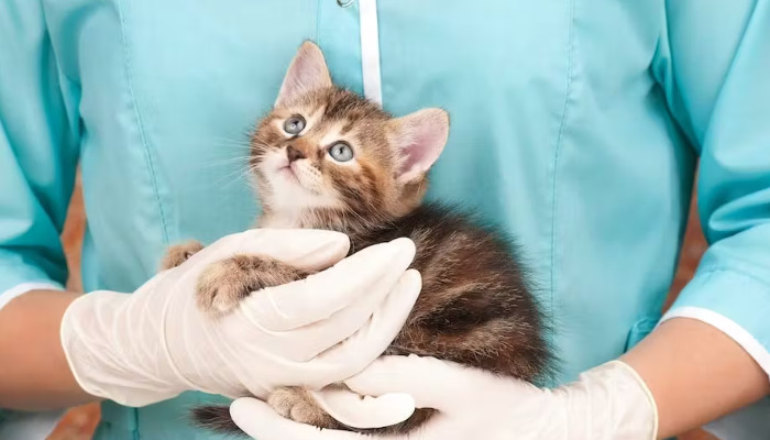
Diagnosing dry FIP begins with a thorough clinical history and physical exam. Most affected cats are young, often under two years of age, and share an environment with other cats. Chronic weight loss, intermittent fever unresponsive to antibiotics, lethargy, anorexia, and sometimes jaundice are common. Ocular and neurologic manifestations—such as uveitis, anisocoria, behavioral changes, or seizures—may also surface.
On palpation, cats may display abdominal discomfort, organomegaly (most often enlarged kidneys or lymph nodes), or signs of icterus. However, none of these are unique to dry FIP; many other diseases—including lymphoma, toxoplasmosis, and bacterial infections—present similarly.
Laboratory Testing: Hematologic and Biochemical Clues
Routine bloodwork can raise suspicion but not confirm dry FIP. Characteristic findings include:
Lymphopenia and neutrophilia
Non-regenerative anemia
Hyperglobulinemia, usually with a low albumin:globulin ratio (<0.8 is highly suspicious).
Elevated liver enzymes or hyperbilirubinemia
Azotemia when kidneys are involved
Serum protein electrophoresis may indicate polyclonal gammopathy, which frequently occurs with FIP but can also be seen in other inflammatory conditions.
Critically, none of these abnormalities are pathognomonic for dry FIP—they simply help place the disease higher on the list of differentials.
Feline Coronavirus Antibody Titers
Testing for feline coronavirus antibody titers can be informative, but it is fraught with ambiguity. High antibody levels suggest exposure, but not necessarily FIP, since many clinically healthy cats have robust titers.
A positive FCoV titer alone cannot distinguish FIP from benign coronavirus exposure. However, in combination with clinical and lab findings, high titers may tip the diagnostic scale further toward FIP—especially in symptomatic, young cats.
Advanced Imaging: Ultrasound and MRI
Imaging technology helps fill the gap left by the absence of effusive fluid. Abdominal ultrasound, in skilled hands, can identify structural abnormalities linked to dry FIP. Such findings include:
Enlarged, irregular kidneys, often with mottled echogenicity
Enlarged mesenteric lymph nodes
Granulomatous lesions in liver, spleen, or intestines
Thickened intestinal walls with loss of normal architecture
MRI, although less commonly available, may be especially useful for neurologic or ocular FIP, delineating granulomatous changes, meningeal enhancement, or subtle masses.
Imaging alone rarely definitively proves FIP, but when coupled with typical clinical findings and laboratory results, it adds strong circumstantial evidence.
Cytology and Fine Needle Aspirates
In the absence of an effusion, cytology can still provide useful clues. Ultrasound-guided aspirate of enlarged lymph nodes, kidneys, liver, or affected tissues may reveal macrophage-dominated, pyogranulomatous inflammation, sometimes with necrosis.
While these findings suggest FIP, they also occur in other diseases. Cytologic analysis must be interpreted in context and cannot replace the definitive value of full-thickness tissue biopsies and IHC.
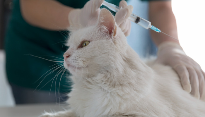
Molecular Diagnostics: FCoV PCR and Genetic Testing
Polymerase chain reaction (PCR) testing for FCoV RNA on blood, tissue, or aspirates has become an increasingly popular diagnostic adjunct. While PCR can detect viral RNA, a positive result is not synonymous with FIP since non-pathogenic coronaviruses are common in cats.
Some laboratories offer PCR panels designed to identify “FIP-type” mutations (spike gene mutations such as M1058L or S1060A). These mutations are enriched in FIP tissues but not exclusive, and a negative result does not rule out the disease.
Combining PCR with clinical suspicion strengthens the diagnostic case, but results must be interpreted cautiously.
Alpha-1 Acid Glycoprotein (AGP) and Other Acute Phase Proteins
AGP is a major acute-phase protein that often rises markedly in cats with FIP. Very high AGP levels (>1.5 mg/mL) are strongly associated with the disease; however, they also elevate in other inflammatory or neoplastic conditions.
AGP may be used as a supportive test. Its application is best as part of a multi-modal diagnostic strategy, not in isolation.
Ocular and Neurologic Evaluation
FIP frequently targets the eyes and central nervous system, especially in dry cases. Slit-lamp examination may reveal anterior uveitis, keratic precipitates, or retinal granulomas. Tonometry may show increased intraocular pressure.
Neurologically, FIP can produce signs ranging from intermittent ataxia to seizures, cranial nerve deficits, or nystagmus. Cerebrospinal fluid (CSF) analysis sometimes uncovers elevated protein or mononuclear pleocytosis, though these are not specific to FIP.
If ocular or CNS involvement is prominent, clinicians may collect aqueous humor or CSF for PCR or cytology. Again, while supportive, none of these approaches are definitive.
Diagnostic Algorithms and Scoring Systems
To sharpen the diagnostic stake, various researchers have developed FIP scoring systems. These algorithms assign points to features such as age, breed, exposure risk, clinical signs, lab results, imaging findings, and AGP levels. When the cumulative score reaches a threshold, the likelihood of FIP—particularly dry forms—rises sufficiently to justify treatment or prognostic counseling.
Examples include the Furr & Kipar FIP Diagnostic Algorithm and the Rivalta Test Algorithm (although the Rivalta test pertains more to wet FIP).
Such structured approaches do not substitute for biopsy, but help clinicians integrate multiple lines of evidence and reduce diagnostic uncertainty.
The Role of Response to Therapy
With the advent of effective antiviral therapies, notably GS-441524 and related compounds, some veterinarians have used a cat’s clinical improvement on FIP-specific treatment as retrospective confirmation. While ethically problematic (as some cats may improve with aggressive supportive care alone), this “therapeutic trial” approach has become more common.
Cats with suspected dry FIP who dramatically improve after a course of antivirals likely did suffer from FIP. Nonetheless, reliance on therapeutic response alone for diagnosis may cause diagnostic confusion or mislead owners—especially in the context of ambiguous clinical signs.
Genetic Predisposition and Breeder Guidance
Dry FIP tends to disproportionately affect young, purebred cats—Birmans, Bengals, and Ragdolls are repeatedly cited. Breeders can help lower FIP risk by reducing overcrowding, segregating kittens from adults with unknown FCoV status, and rigorous hygiene. Yet, even in best-case scenarios, FCoV can become endemic, complicating diagnosis further.
Emerging Non-Invasive Technologies
Current veterinary research includes leveraging next-generation sequencing, advanced proteomics, and even artificial intelligence algorithms to distinguish FIP from lookalike conditions. These technologies, while not widely available, suggest a future in which non-invasive testing may provide highly specific, rapid answers—even for dry FIP.
Ethical Considerations
In practice, feline clinicians must balance the need for diagnostic certainty with the welfare of their patients. Pursuing invasive tests when the likelihood of benefit is low or when the risk is high is ethically questionable. Veterinary consensus in the United States increasingly supports a “weight of evidence” approach, using non-invasive modalities to reach a presumptive diagnosis when biopsy is impractical or unsafe.
Key Points and Clinical Pearls
Dry FIP can infrequently be definitively diagnosed without tissue biopsy.
Careful synthesis of age, history, clinical signs, lab findings, imaging, antibody titers, AGP levels, and molecular tests can yield very strong presumptive evidence.
Absence of pathognomonic results means that diagnosis remains probabilistic—but often sufficiently robust to guide humane decision-making or justify antiviral therapy.
Ongoing research may soon improve non-invasive test accuracy.
Reference List
1. Kipar, A., & Meli, M. L. (2014). Feline infectious peritonitis: still an enigma? Veterinary Pathology, 51(2), 505-526.
2. Felten, S., & Hartmann, K. (2019). Diagnosis of feline infectious peritonitis: a review of current literature. Viruses, 11(11), 1068.
3. Addie, D. D., & Jarrett, O. (1992). Feline coronavirus infections. British Veterinary Journal, 148(3), 223-254.
4. Tasker, S. (2018). Diagnosis of feline infectious peritonitis: update on evidence supporting laboratory testing and future developments. The Veterinary Journal, 2018 Jun;238:31-38.
5. Stranieri, A., et al. (2018). Diagnostic markers for effusive and non-effusive feline infectious peritonitis. Veterinary Clinical Pathology, 47(2), 272-282.
6. Pedersen, N. C. (2009). Feline infectious peritonitis virus infection. Feline Infectious Diseases (pp. 73-88).
7. Dempsey, S. M., & Ewing, P. J. (2011). A review of the pathogenesis, clinical diagnosis, and management of feline infectious peritonitis. Journal of Feline Medicine and Surgery, 13(7), 377-387.
8. Fischer, Y., et al. (2011). Detection of feline coronavirus spike gene mutations as a diagnostic tool for feline infectious peritonitis. Journal of Feline Medicine and Surgery, 13(10), 803-807.
9. Paltrinieri, S., et al. (2007). Serum protein electrophoresis and acute phase protein measurement for diagnosis of feline infectious peritonitis. Veterinary Clinical Pathology, 36(2), 133-138.
10. Riemer, F., et al. (2016). Clinical and laboratory features of cats with feline infectious peritonitis–a retrospective study of 231 confirmed cases (2000–2010). Journal of Feline Medicine and Surgery, 18(4), 348-356.
11. Addie, D. D., et al. (2009). Feline infectious peritonitis: ABCD guidelines on prevention and management. Journal of Feline Medicine and Surgery, 11(7), 594-604.