Are Diagnostic Criteria Different for Various Types of FIP

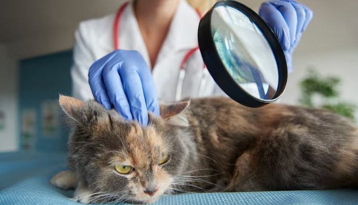
Feline Infectious Peritonitis (FIP) stands among the most perplexing and devastating diseases to affect domestic cats. The diagnostic process is particularly challenging due to the disease’s varied clinical manifestations and the absence of a single definitive test. As research advances and understanding grows, veterinarians and feline owners continue to ask: Are the diagnostic criteria different for various types of FIP? This article explores the array of FIP types, compares diagnostic approaches, examines the underlying reasons for these differences, and offers insight into the complexities of this disease, integrating scientific findings and practical perspectives.
FIP Overview and Its Clinical Diversity
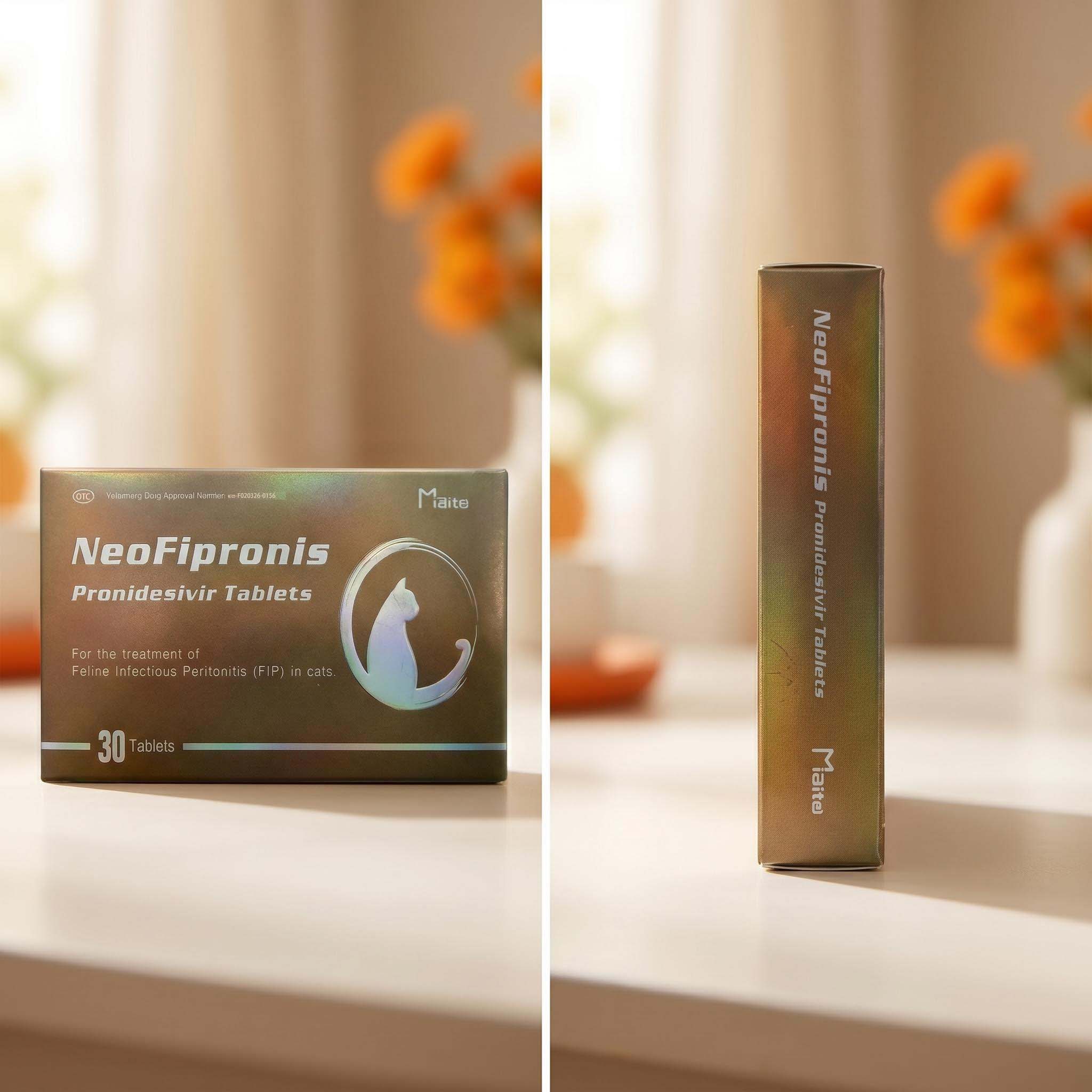
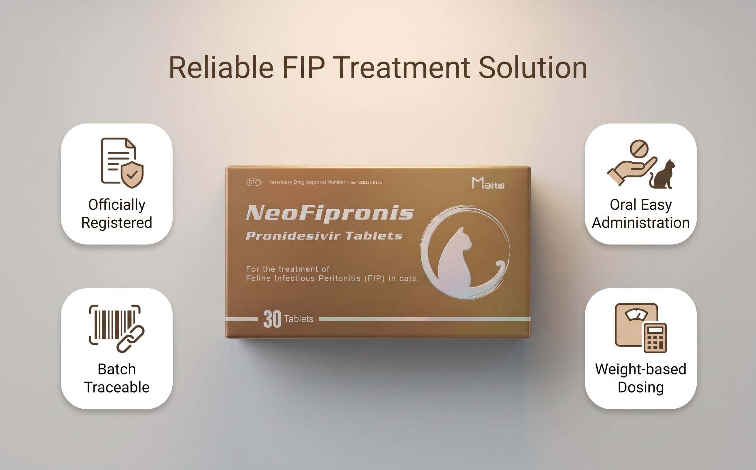
FIP is caused by a mutated form of feline coronavirus (FCoV) that triggers a systemic, often fatal immune-mediated response. Not all FCoV-infected cats develop FIP; most experience mild, transient symptoms or remain asymptomatic. However, when the virus mutates within the host, it results in FIP, which is invariably fatal without aggressive intervention.
Clinical signs vary widely and are classically divided into two major forms:
'Effusive' (wet) FIP: Characterized by fluid accumulation in body cavities.
'Non-effusive' (dry) FIP: Marked by granulomatous lesions without significant fluid build-up.
Within these, subtypes exist, including ocular and neurological presentations, further complicating diagnostic strategies.
Wet vs. Dry FIP: Hallmark Differences
The distinction between wet and dry FIP forms underpins much of the variation in diagnostic criteria.
Wet FIP: The classic effusive form typically features significant effusion in the abdomen, chest, or both. Clinical signs include abdominal distension, breathing difficulties, lethargy, poor appetite, and fever unresponsive to antibiotics.
Dry FIP: In contrast, non-effusive FIP presents less obviously with vague signs such as persistent fever, weight loss, jaundice, and organ enlargement. Neurological and ocular involvement may dominate the clinical picture.
Diagnostic Gold Standards and Their Limitations
A major challenge in FIP diagnosis is the lack of a pathognomonic test. Existing assays detect antibodies, viral RNA, or antigens, but none can reliably differentiate FIP from self-limiting FCoV infection.
Laboratory Findings
Laboratory diagnostics provide clues but rarely definitive answers. Common findings across both forms include:
Hyperglobulinemia (increased globulin levels)
Lymphopenia
Anemia
Elevated protein levels in effusions or serum
However, these changes are not unique to FIP and occur with other diseases.
Effusive FIP: Diagnostic Emphasis
For wet FIP, analysis of effusions is central:
Rivalta test: A positive Rivalta test supports FIP, but false positives and negatives occur.
Effusion analysis: Straw-colored, viscous, protein-rich fluid with low cellularity and high total protein (>3.5 g/dL) raises suspicion.
Molecular testing (PCR) can detect FCoV RNA in effusions, confirming infection but not necessarily FIP.
Dry FIP: Diagnostic Pathways
Dry FIP diagnosis relies heavily on imaging and tissue analysis:
Imaging: Ultrasound or radiographs may reveal organ enlargement, lymphadenopathy, or focal masses.
FNA/Biopsy: Fine-needle aspirates or tissue biopsies provide cellular detail. Immunohistochemical staining for FCoV antigen within macrophages is highly suggestive.
Neurological and ocular involvement necessitates advanced diagnostic imaging (MRI, CT) and may require cerebrospinal fluid (CSF) or ocular fluid analysis.
Ocular and Neurological FIP: Specialized Considerations
The 'atypical' forms—ocular and neurological FIP—pose unique diagnostic challenges and often overlap with the dry type.
Ocular FIP: Presents with anterior uveitis, retinal detachment, and aqueous flare. Ophthalmologic examination, along with aqueous humor analysis, can suggest FIP but is rarely definitive.
Neurological FIP: May cause seizures, ataxia, and behavioral changes. CSF analysis can reveal elevated protein and mononuclear pleocytosis, though these features overlap with other meningoencephalitides.
Confirmation in both cases may depend on ante-mortem tissue sampling and immunohistochemistry, which is invasive and often declined in clinical practice.
Serologic and Molecular Testing: Strengths and Weaknesses
Serologic testing for FCoV antibodies reveals exposure but fails to differentiate FIP from benign FCoV infection. High antibody titers and PCR detection of FCoV RNA, especially in tissue samples or effusions, lend support but do not confirm FIP due to the prevalence of FCoV in the feline population.
Newer molecular assays (e.g., Real-Time PCR) targeting specific mutations in the S gene associated with the FIP-causing strain show promise, especially when performed on tissue biopsies. However, these are not universally available and may provide false negatives if sampling is suboptimal.
Histopathology: The Definitive Answer
When feasible, histopathological tissue analysis remains the gold standard.
Wet FIP: Necrotizing vasculitis with fibrinous exudation in affected organs.
Dry FIP: Pyogranulomatous inflammation with macrophage infiltration.
Immunohistochemical staining for FCoV antigen within lesion macrophages clinches the diagnosis.
Despite its definitive nature, biopsies are rarely performed ante-mortem due to invasiveness and clinical risk.
Age and Breed Predilections Affect Diagnostic Approach
FIP most commonly affects young cats (under 2 years), shelter cats, and certain breeds (e.g., Bengals, Abyssinians, Birmans). Higher suspicion in these populations may lower the diagnostic threshold.
Differential Diagnoses and Confounding Factors
Similar presentations occur in diseases such as lymphoma, bacterial peritonitis/pleuritis, toxoplasmosis, and hepatic lipidosis, particularly with dry FIP. Comorbidities further cloud the clinical picture, demanding a careful and comprehensive diagnostic work-up.
Are Criteria Truly Different?
While the goal—to confirm FIP—is constant, the means shift according to type:
Wet FIP relies on effusion analysis, serology, and PCR from fluids.
Dry FIP pushes for tissue imaging, cytology, and, when possible, biopsy with immunohistochemistry.
Ocular and neurological FIP demand ocular/neurological fluid analysis and advanced imaging, with tissue confirmation as a gold standard.
These variations derive not only from the clinical signs but also from technical limitations, owner preferences, and the invasiveness of certain tests.
Emerging Approaches and Their Impact
In recent years, advances in molecular diagnostics—such as RT-PCR and genetic sequencing—have improved sensitivity, especially for detecting FIP-associated mutations. Point-of-care tests are evolving, though none are yet considered definitive for every presentation.
Therapeutic response to experimental FIP antivirals (e.g., GS-441524) is sometimes invoked as indirect confirmation, with dramatic clinical improvement taken to support an FIP diagnosis. However, this is not standard protocol.
Integrating Findings: A Clinician’s Strategy
Veterinarians must adapt diagnostic protocols to the cat’s presentation and owner circumstances. A thorough patient history—age, breed, environment, and disease course—guides differential diagnosis. Laboratory and imaging findings must be interpreted in the full clinical context, never in isolation.
The decision to pursue invasive diagnostics (e.g., tissue biopsy) balances medical necessity and animal welfare. Many clinicians use a combination of strongly suggestive findings—persistent fever, signalment, effusive features, high coronavirus titers, and supportive imaging—to make a ‘presumptive diagnosis’ when definitive proof is unattainable.
Conclusions Drawn from Clinical Practice
Diagnostic criteria do differ between FIP types. Clinical presentation, affected organs, and sample accessibility drive the choice and interpretation of tests. Ongoing research into improved diagnostics and understanding FCoV pathogenesis continues to shape the future of FIP recognition and management.
References
1. Kipar, A., & Meli, M. L. (2014). Feline infectious peritonitis: Still an enigma? Veterinary Pathology, 51(2), 505–526. https://doi.org/10.1177/0300985814522077
2. Hartmann, K. (2005). Feline infectious peritonitis. Veterinary Clinics of North America: Small Animal Practice, 35(2), 39–79. https://doi.org/10.1016/j.cvsm.2004.10.011
3. Felten, S., & Hartmann, K. (2019). Diagnosis of feline infectious peritonitis: A review of backgrounds and current advances. Veterinary Clinical Pathology, 48(2), 273–284. https://doi.org/10.1111/vcp.12701
4. Pedersen, N. C. (2014). An update on feline infectious peritonitis: Diagnostics and therapeutics. Veterinary Journal, 201(2), 133–141. https://doi.org/10.1016/j.tvjl.2014.04.017
5. Addie, D. D., et al. (2020). Progress in diagnosing and managing feline infectious peritonitis. The Veterinary Record, 186(8), 254–256. https://doi.org/10.1136/vr.m963
6. Tasker, S. (2018). Diagnosis of feline infectious peritonitis: Update on evidence supporting laboratory testing. Journal of Feline Medicine and Surgery, 20(3), 228–243. https://doi.org/10.1177/1098612X18758592
7. Stranieri, A., et al. (2023). Molecular diagnostics for feline infectious peritonitis: Emerging technologies. Veterinary Microbiology, 281, 109673. https://doi.org/10.1016/j.vetmic.2022.109673
8. Giori, L., et al. (2011). Feline coronaviruses: Pathogenesis of FIP and molecular diagnostics. Diagnostic Microbiology and Infectious Disease, 71(2), 153–160. https://doi.org/10.1016/j.diagmicrobio.2011.06.014
9. Riemer, F., et al. (2016). Clinical and laboratory features of FIP—How to improve the diagnostic approach. Journal of Feline Medicine and Surgery, 18(2), 108–119. https://doi.org/10.1177/1098612X15596338
10. Barker, E. N., et al. (2017). Ocular and neurological forms of feline infectious peritonitis: Diagnostic challenges and approaches. Veterinary Ophthalmology, 20(S1), 7–15. https://doi.org/10.1111/vop.12264