What Lab Indicators Matter Most During FIP Treatment
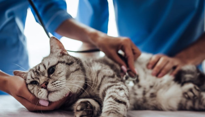
Feline Infectious Peritonitis (FIP) stands among the most daunting challenges in feline internal medicine. An immune-mediated, often fatal condition caused by specific mutations in feline coronavirus (FCoV), FIP once meant inevitable loss, but recent advances in antiviral therapy have offered new hope. Today's clinicians and pet owners are tasked with careful monitoring, not only of clinical signs but especially of key laboratory values that reveal the underlying health status, response to treatment, and prognosis.
Understanding which laboratory indicators to track, what changes signify, and how these correlate with a cat's progress is critical for effective FIP management. For newly diagnosed cases, those under active treatment (such as with GS-441524 or GC376), and cats in remission, a comprehensive grasp of relevant biomarkers is pivotal. This article compiles current clinical knowledge on lab indicators that matter most during FIP treatment.
Complete Blood Count (CBC): Interpreting the Basics and Beyond
The CBC is typically the first stop in feline diagnostics. Anemia (usually non-regenerative normocytic, normochromic) is a hallmark of chronic inflammation in FIP. Successful treatment frequently brings gradual normalization of red blood cell counts.
Leukocytosis—an increased white blood cell (WBC) count—reflects the body's inflammatory response. Neutrophilia, particularly with a left shift (immature neutrophils), often accompanies active FIP. As treatment progresses, resolution or reduction in WBC count can suggest therapeutic efficacy. Conversely, leukopenia may result from bone marrow suppression, sometimes due to the disease itself or rarely from certain antiviral treatment side effects.
Thrombocytopenia (low platelets), observed in many FIP cats, may stem from immune-mediated destruction or disseminated intravascular coagulation. Restoration of normal platelet levels is a positive response indicator. Thus, trending CBC values over time assists clinicians in gauging both disease progression and treatment response.
Serum Biochemistry: Liver and Kidney Health Under the Spotlight
Serum chemistry panels shed light on organ function and the severity of systemic involvement, crucial given FIP’s tendency toward multisystemic disease.
Hyperglobulinemia, especially elevated gamma globulins, is near-universal in FIP, reflecting chronic immune stimulation. As cats respond to treatment, total globulin concentrations frequently decline, sometimes lagging behind clinical improvement. Albumin, a negative acute-phase protein, often decreases in severe FIP. The albumin/globulin (A/G) ratio is a cardinal metric: ratios below 0.6 are suspicious for FIP, and rising ratios can denote successful therapy.
Elevations in liver enzymes (ALT, AST) and bilirubin suggest hepatic involvement—a grave prognostic sign historically. However, many cats now recover hepatic function with proper antiviral therapy. Similarly, renal function markers (BUN, creatinine) require monitoring, especially in cats with non-effusive neurological or ocular FIP.
Electrolyte imbalances (hyponatremia, hypokalemia) may reflect severe effusive disease, dehydration, or organ failure. Serial monitoring and correction are critical during the acute phase of treatment.
Acute Phase Proteins and Inflammatory Markers: CRP, SAA, AGP
C-reactive protein (CRP) and Serum Amyloid A (SAA) are acute-phase reactants, increasingly used to track inflammation in FIP. Elevated CRP and SAA at diagnosis often correlate with disease severity. Rapid reduction after initiation of antiviral drugs (e.g., GS-441524) indicates favorable response.
Alpha-1 acid glycoprotein (AGP) is less conventionally measured but has particular utility in distinguishing FIP from other conditions. Its marked elevation tends to subside as cats improve.
These biomarkers are not infallible—false positives and negatives occur—but together, they illuminate disease trajectory when interpreted alongside clinical signs and other lab values.
Effusion Analysis: The Gold Standard for Wet FIP
Effusive ("wet") FIP is characterized by protein-rich fluid accumulation in the abdomen or thorax. Fluid analysis remains highly diagnostic:
High protein concentration (>3.5 g/dL), especially exudates with low cell counts, supports FIP diagnosis.
Rivalta test positive results, though non-specific, are highly suggestive in suspect cases.
Cytology often reveals non-septic neutrophils, macrophages, and proteinaceous background.
Serial analysis of effusion is less common but can monitor resolution. Gradual reduction or complete disappearance typically mirrors major improvement.
Coronavirus-Specific Testing: PCR, Serology, and Antigen Detection
Reverse-transcriptase PCR (RT-PCR) testing on blood, tissue, or effusion can confirm FCoV RNA, though negative results do not exclude disease due to intermittent viremia. PCR on effusion fluid provides the best diagnostic yield. Many clinicians combine PCR with FCoV antibody titers—high titers may support FIP, but overlap with benign enteric FCoV limits specificity.
During treatment, decreasing RT-PCR copy numbers are encouraging but not foolproof for predicting remission. Residual viral RNA may persist even after clinical cure; thus, PCR should complement, not supplant, traditional lab monitoring.
Immunocytochemistry and Immunohistochemistry: Cellular Insights
For challenging cases, immunocytochemistry (ICC) or immunohistochemistry (IHC) can demonstrate FCoV antigen within macrophages from effusion fluid or tissue biopsies, providing near-definitive diagnostics. In therapy, repeat biopsies are rare, but declining antigen presence can support recovery.
Prognostic Indicators: What Predicts Survival and Remission?
When evaluating a newly diagnosed FIP cat, certain labs best predict outcome and should be monitored closely:
Baseline A/G ratio: Higher ratios predict better survival.
CRP/SAA trajectory: Rapid declines correlate with clinical response.
Platelet count: Recovery of platelets predicts remission.
Serum bilirubin: Persistent elevation portends poor outcome.
Hematocrit recovery: Anemia resolution signals improvement.
Resolution of effusion: Nearly always required for prolonged survival in wet FIP.
No single marker gives all answers. Instead, integrating several laboratory indicators, repeating them serially, and cross-referencing against clinical signs leads to informed decisions.
Monitoring Treatment Response: Frequency and Strategy
Clinicians generally advise standardized lab monitoring at key intervals:
Baseline: Full CBC, chemistry, CRP/SAA, effusion analysis, PCR.
Every 2–4 weeks: CBC, chemistry, CRP/SAA, effusion status.
After treatment completion: CBC and chemistry to confirm stability and absence of relapse.
Cats experiencing side effects (anorexia, vomiting, jaundice, neurological changes) may need more frequent monitoring. Owners informed about what each test represents and how indicators may fluctuate are better prepared to make decisions in partnership with their veterinary team.
Special Considerations in Neurological and Ocular FIP
Cats with neurological or ocular FIP present unique diagnostic and therapeutic challenges. Cerebrospinal fluid (CSF) analysis becomes critical:
CSF protein concentration: Mild to marked elevations are common.
CSF cell count: Mononuclear pleocytosis (increased cells) indicates meningoencephalitis.
FCoV PCR in CSF: Sometimes positive; presence supports diagnosis.
Serial CSF monitoring, while invasive, can assess treatment response in select cases.
Ocular FIP may benefit from periodic measurement of intraocular proteins, but clinical assessment (fundoscopy, response to therapy) takes precedence.
Potential Laboratory Side Effects of Antiviral Therapy
Most FIP therapy today involves nucleoside analogues, which are generally well-tolerated but may cause mild elevations in liver enzymes, occasional cytopenias, or rare kidney effects. Monitoring for these helps ensure safe and effective therapy.
Supportive care—hydration, antiemetics, nutritional support—may also impact lab markers (e.g., higher glucose or corrected electrolytes), underlining the importance of context.
Integrating Laboratory and Clinical Signs: A Holistic Approach
Laboratory indicators rarely operate in isolation. Fever, weight gain, return of appetite, reduction in effusion, and improved attitude should always be integrated with lab findings. Sometimes, lab normalization trails behind clinical recovery, meaning patience and thorough monitoring are necessary.
Conversely, worsening labs (increased bilirubin, persistent anemia, declining A/G ratio, rising CRP/SAA) may precede clinical relapse, prompting early intervention.
Laboratory Values in the Post-Treatment and Remission Period
Even after treatment, cats remain at risk of FIP recurrence. Periodic labs—at least every 2–3 months initially—are wise, especially in cats previously showing severe lab derangements. Owners should watch for new clinical signs and seek repeat testing promptly.
Some clinicians advocate for a gradual reduction in the frequency of monitoring after 6–12 months in complete remission, provided all labs remain stable and the cat is clinically healthy.
Lab Monitoring Recommendations for Veterinarians and Cat Owners
Successful FIP management is a partnership—veterinarians bring expertise in lab interpretation, owners bring intimate knowledge of their cat’s daily habits. Labs to track closely include:
CBC: Focus on anemia, WBC, platelets.
Chemistry panel: Albumin, globulin, liver/kidney markers, electrolytes.
CRP/SAA: Trend acute-phase reactants.
Effusion protein/cell count: Where relevant.
PCR (if available): FCoV RNA titers.
All results are best interpreted over time rather than in isolation, with awareness of typical patterns, possible side effects from drugs, and expected fluctuations.
References
1. Pedersen, N.C. (2019). The History of FIP and the Discovery of Effective Antiviral Treatments. J Feline Med Surg, 21(9): 809-818.
2. Dempsey, S., & Ewing, P.J. (2011). Feline Infectious Peritonitis: Diagnostic Dilemmas and Therapeutic Options. Vet Clin North Am Small Anim Pract, 41(2): 345-364.
3. Stranieri, A., et al. (2021). Laboratory Abnormalities in Cats with Feline Infectious Peritonitis. Animals, 11(2): 353.
4. Tasker, S. (2018). Diagnosis of Feline Infectious Peritonitis: Update on Evidence Supporting Laboratory Testing. J Feline Med Surg, 20(3): 228-243.
5. Giori, L., Giordano, A., Giudice, C., et al. (2011). Performance of a Commercially Available Immunochromatographic Test for the Detection of Feline Coronavirus Antigen. Vet J, 190(2): 348-354.
6. Diaz, C., et al. (2024). Monitoring of Acute Phase Proteins During GS-441524 Treatment of FIP. Vet Microbiol, 285: 109987.
7. Felten, S., et al. (2020). Correlation of Albumin/Globulin Ratio and Survival Time in Cats with FIP Treated with Nucleoside Analogues. J Feline Med Surg, 22(4): 302-312.
8. Murphy, B.G., et al. (2018). Treatment of FIP with GC376 and Monitoring Laboratory Values. Vet Pathol, 55(5): 755-768.
9. Addie, D.D., et al. (2022). International Field Experience with FIP Diagnosis and GS-441524 Therapy. Viruses, 14(1): 123.
10. Hartmann, K. (2020). Feline Infectious Peritonitis—Recent Knowledge about Diagnostics and Therapy. Tierarztl Prax Ausg K Kleintiere Heimtiere, 48(2): 74-83.